Japanese
English
- 有料閲覧
- Abstract 文献概要
- 1ページ目 Look Inside
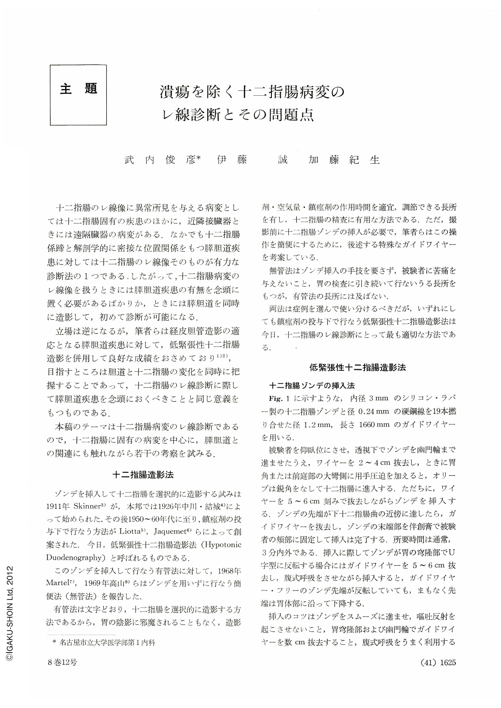
十二指腸のレ線像に異常所見を与える病変としては十二指腸固有の疾患のほかに,近隣接臓器ときには遠隔臓器の病変がある.なかでも十二指腸係蹄と解剖学的に密接な位置関係をもつ膵胆道疾患に対しては十二指腸のレ線像そのものが有力な診断法の1つである.したがって,十二指腸病変のレ線像を扱うときには膵胆道疾患の有無を念頭に置く必要があるばかりか,ときには膵胆道を同時に造影して,初めて診断が可能になる.
立場は逆になるが,筆者らは経皮胆管造影の適応となる膵胆道疾患に対して,低緊張性十二指腸造影を併用して良好な成績をおさめており1)2),目指すところは胆道と十二指腸の変化を同時に把握することであって,十二指腸のレ線診断に際して膵胆道疾患を念頭におくべきことと同じ意義をもつものである.
We are of the opinion that hypotonic duodenography is the method of choice in the diagnosis of lesions in the duodenum, and in this paper is introduced our procedure. Also is decribed a guide wire for facilitating the introduction of duodenal probe together with the gist of probe insertion utilizing the wire.
The essentials of x-ray diagnosis of lesions in the duodenum are also presented after dividing them into functional abnormalities, congenital ones, diverticulum, neoplasms, inflammation and others.
As was proposed by Dr. Hayashi of the Department of Pathology in our College of Medicine, the papillary region is defined as that conical area within the duodenal lumen where choledochopancreatic duct is surrounded with the sphincter of Oddi. Hayashi assumed that papillary cancer mostly arises from the bile duct within the duodenal wall, but from the viewpoint of roentgenologic diagnosis we believe that papillary cancer should be considered as a lesion of the duodenum.
In x-ray diagnosis of cancer and other lesions of the papillary region, simultaneous coemployment of hypotonic duodenography and percutaneous transhepatic cholangiography is most effective.
A case of carcinoma in the minor papillaiy illustrated as well, with some comments on the necessity in future of many-sided investigations regarding the accesory papilla. Finally, some problems in dealing with duodenal lesions have been discussed.

Copyright © 1973, Igaku-Shoin Ltd. All rights reserved.


