Japanese
English
- 有料閲覧
- Abstract 文献概要
- 1ページ目 Look Inside
はじめに
隆起性病変の中央部に浅い潰瘍を認め,その噴門側にⅡc様病変が存在した特異な1症例を経験した.術前Ⅲ+Ⅱcを疑ったが,粘膜下腫瘍,またはⅢ型の進行癌も否定できず,確診が得られぬままに手術を施行した.術後Ⅱc+Ⅲ型早期胃癌に限局性のリンパ細網組織の増生が重なった症例であったので報告する.
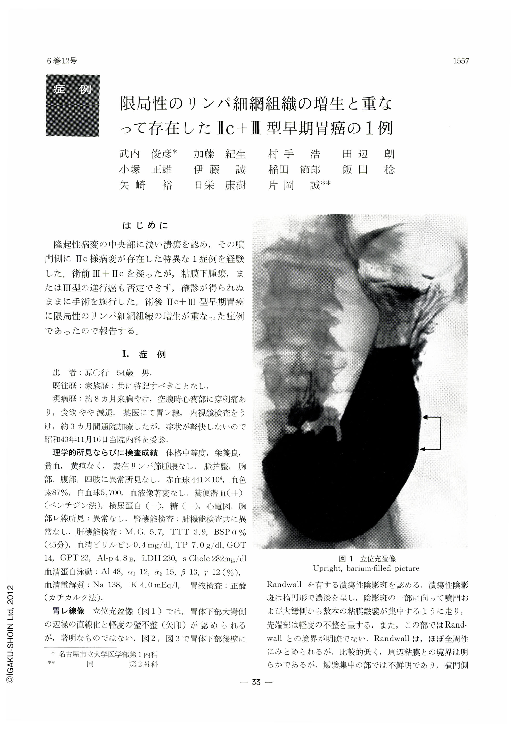
A 54-year-old man visited the authors' clinic complaining of epigastric pain. X-ray study of the stomach revealed on the posterior wall of the lower body a barium shadow fleck suggesting of an ulcer with distinct marginal borders extending almost in its whole circumference, although slightly ill-defined on the cardiac side. The lesion was visualized by endoscopy at the same site as a relatively shallow ulcer with irregular margins. The ulcer with its surrounding area was elevated as a whole, and elevations gradually and gently merged into the encircling normal mucosa. Contiguous to the lesion, its oral mucosa was discolored in some parts looking as if cancer infiltration had extended there. Gross specimen of the removed stomach disclosed at a site about 7cm oral from the pyloric ring a relatively deep ulcer, measuring 10×10 mm, neighbored still on its oral side by a shallow depressed lesion which measured 15×10 mm. Histologically, a localized tumor formation (adenocarcinoma acinosum), measuring 10 mm in its greatest diameter and localized within the submucosa, was seen in the site corresponding to the deep ulcer. Tumor cells and lymphoreticular hyperplasia were well intermixed. The adjacent lesion on the oral side, a shallow depression, was tubular adenocarcinoma localized within the mucosa. The whole picture constituted Ⅱc+Ⅲ type early gastric cancer coexistent with localized lyrnphoreticular hyperplasia. In Japan so far only four such cases including the present one have been reported. As histologically this lesion shows a structure to be called “medullary carcinoma with lymphoid stroma”, prognosis of this variety is considered favorable. Finally, reference has been made to a possible development pattern of the lesion.

Copyright © 1971, Igaku-Shoin Ltd. All rights reserved.


