Japanese
English
- 有料閲覧
PPV購入案内
1,320円 (1,200円+税10%) こちらは、433ページから438ページの文献です。購入には医書.jp本会員登録が必要です。
会員登録完了後に改めて上記「購入サイトへ」を選択してください。
または「購入サイトへ」選択後に「購入ログイン」、「新規会員登録」の順に進んでください。
会員登録について詳しくはをご確認ください。
- Abstract 文献概要
- 1ページ目 Look Inside
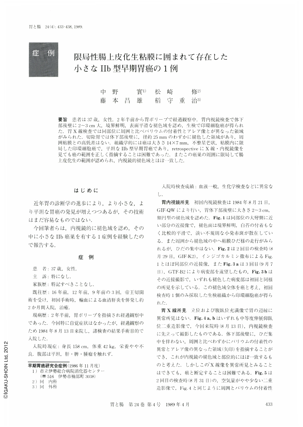
要旨 患者は37歳,女性.2年半前から胃ポリープで経過観察中.胃内視鏡検査で体下部後壁に2~3cm大,境界鮮明,表面平滑な褪色域を認め,生検で印環細胞癌が得られた.胃X線検査では同部位に周囲と比ベバリウムの付着性とアレア像とが異なった領域がみられた.切除胃では体下部後壁に,径約25mmのわずかに褪色した領域があり,周囲粘膜との高低差はない.組織学的には癌は大きさ14×7mm,不整星芒状,粘膜内に限局した印環細胞癌で,平坦なⅡb型早期胃癌であり,retrospectiveにX線・内視鏡像を見ても癌の範囲を正しく指摘することは困難であった.またこの癌巣の周囲に限局して腸上皮化生の範囲が認められ,内視鏡的褪色域とほぼ一致した.
A 37-year-old asymptomatic woman who had been told to have a gastric polyp in the antrum about two and a half years before. She visited our hospital on Aug. 13, 1984, to have annual gastric examination.
Endoscopic examination revealed a round, sharply demarcated discolored area, about 2 to 3 cm in size on the posterior wall of the lower body. The surface of this area was smooth and had slightly reddish flecks (Figs. 1-3). Biopsy specimen taken from this area revealed signet-ring cell carcinoma. X-ray examination of the stomach also showed irregular mucosal pattern and slightly abnormal barium coating on the posterior wall of the lower body (Figs. 4 and 5). Consequently, subtotal gastrectomy was performed on Sept. 11, 1984.
Resected specimen of the stomach had a subtle discolored area with neither depression nor elevation on the posterior wall of the lower body (Fig. 6).
Histologically, signet-ring cell carcinoma was found within the mucosal layer (Figs. 9 and 10). The cancer lesion was 14×7 mm in size and showed neither elevation nor depression, providing diagnostic evidence of type Ⅱb early gastric cancer (Fig. 7). Histological examination also revealed the localized intestinal metaplasia about 25 mm in diameter around this cancer lesion, which corresponded almost exactly with the discolored area (Fig. 7).
Retrospectively, the discolored area noted on endoscopy as well as by macroscopic observation was the localized intestinal metaplasia including the cancer lesion.
Copyright © 1989, Igaku-Shoin Ltd. All rights reserved.