Japanese
English
- 有料閲覧
- Abstract 文献概要
- 1ページ目 Look Inside
- サイト内被引用 Cited by
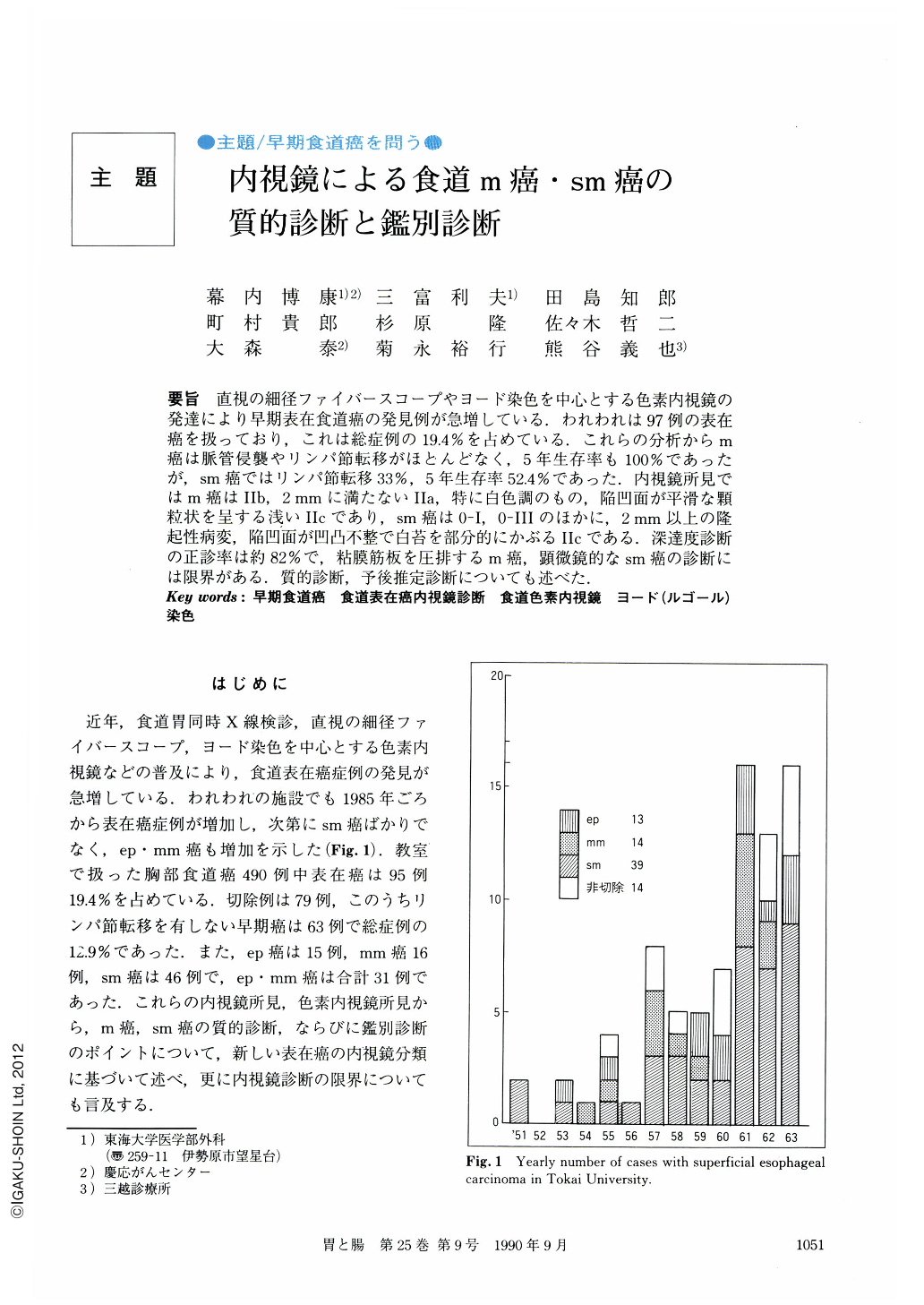
要旨 直視の細径ファイバースコープやヨード染色を中心とする色素内視鏡の発達により早期表在食道癌の発見例が急増している.われわれは97例の表在癌を扱っており,これは総症例の19.4%を占めている.これらの分析からm癌は脈管侵襲やリンパ節転移がほとんどなく,5年生存率も100%であったが,sm癌ではリンパ節転移33%,5年生存率52.4%であった.内視鏡所見ではm癌はⅡb,2mmに満たないⅡa,特に白色調のもの,陥凹面が平滑な顆粒状を呈する浅いⅡcであり,sm癌は0-1,0-Ⅲのほかに,2mm以上の隆起性病変,陥凹面が凹凸不整で白苔を部分的にかぶるⅡcである.深達度診断の正診率は約82%で,粘膜筋板を圧排するm癌,顕微鏡的なsm癌の診断には限界がある.質的診断,予後推定診断についても述べた.
In Japan, detection of patients with early and superficial carcinoma in the esophagus has inproved because of developments of a slender fiberscope, and techniques of chromo-endoscopy such as iodine staining. So far, we have encountered 95 cases with superficial esophageal carcinoma with invasion limited to the submucosal layer. They represent 19.4% of 409 cases with carcinomas in the thoracic esophagus treated in our hospital. Detected analyses of these cases revealed that mucosal carcinomas rarely invade the vessels and metastasize to the lymph nodes. Thus, a 5-year survival rate was 100%. On the other hand, 33% of cases with submucosal carcinoma had lymph node metastasis and their 5-year survival rate was 52.4%. In this paper, we described endoscopic characteristics for their differentiation of mucosal and submucosal carcinoma.
Briefly, mucosal carcinomas are flat (0-Ⅱb), 2 mm or smaller elevated lesions (0-Ⅱa). They often look white, and have shallow erosion (0-Ⅱc) with regular granular bases. On the contrary, submucosal carcinomas are usually elevated lesions larger than 2 mm or deep irregular ulcers covered with white debris. Using these criteria, accuracy in diagnosis was approximately 82%. Some of the mucosal carcinomas invading nearly as far as the mucosal muscle layer and submucosal carcinomas with microscopic invasion of the submucosal layer were misdiagnosed.
Copyright © 1990, Igaku-Shoin Ltd. All rights reserved.