Japanese
English
- 有料閲覧
- Abstract 文献概要
- 1ページ目 Look Inside
はじめに
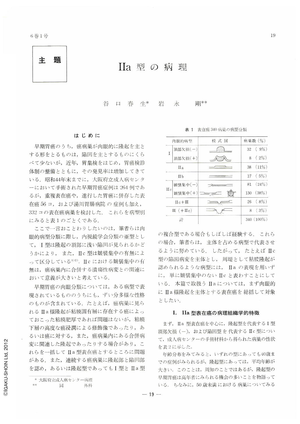
早期胃癌のうち,癌病巣が肉眼的に隆起を主とする形をとるものは,陥凹を主とするものにくらべて少ないが,近年,胃集検をはじめ,胃癌検診体制の整備とともに,その発見率は増加してきている.昭和44年末までに,大阪府立成人病センターにおいて手術された早期胃癌症例は264例であるが,重複表在癌や,進行した胃癌に併存した表在癌56コ,および湯川胃腸病院の症例も加え,332コの表在癌病巣を検討した.これらを病型別にみると表1のごとくである.
ここで一言おことわりしたいのは,筆者らは肉眼的病型分類に際し,内視鏡学会分類の亜型として,Ⅰ型は隆起の頂部に浅い陥凹が見られるかどうかにより,また,Ⅱc型は皺襞集中の有無によって区分している1)2).Ⅱcにおける皺襞集中の有無は,癌病巣内に合併する潰瘍性病変との関連において意義が大きいと考えている.
早期胃癌の肉眼分類については,ある病型で表現されているもののうちにも,ずい分多様な性格のものが含まれている.たとえば,癌病巣に見られるⅡa様隆起が粘膜固有層に存在する癌によっておこった粘膜肥厚であれば問題はないが,粘膜下層の高度な癌浸潤による修飾像であったり,あるいは癌に対する,また,癌病巣内にある合併病変に関連した隆起であったりする場合があり,これらを一括してⅡa型表在癌とするところに問題がある.また,連続する癌病巣に隆起部と陥凹部を認め,あるいは隆起型であってもⅠ型とⅡa型の複合型である場合もしばしば経験する.これらの揚合,筆者らは,主体を占める病型で代表させるように努めている.したがって,たとえばⅡc型の陥凹病変を主体とし,周堤として粘膜隆起が認められるような病型には,Ⅱaの表現を用いずに,単に皺襞集中のないⅡcと表わすことにしている.本篇で取扱うⅡaについては,まず肉眼的にⅡa様隆起を主体とする表在癌を総括して対象としたい.
A histological survey was attempted on 35 lesions of superficial cancer having main macroscopic characteristics to be classified as Ⅱa. In this study depressed type of superficial cancer (Ⅱc) that has marginal elevation was excluded because the authors have regarded it as belonging to a variety of Ⅱc that has no rugal convergency. Excluded as well was Ⅱa-like elevation caused by infiltration and/or proliferation of cancer cell nest in the submucosal level.
Of 35 lesions of grossly Ⅱa type early cancer, 24 were Ⅱa sharply circumscribed from the surrounding mucosa; four had small erosions on their surface; seven were coexistent with adjacent or with Ⅱc within the elevation itself (Ⅱa+Ⅱc); and the rcmaining four were associated with ulcerative change within the Ⅱa lesion.
Regarding depression in the Ⅱa lesion, dell-like one without any surface erosion presented few problems, while erosive depression, either large or small, showed in most instances higher cellular and structural atypism with more submucosal invasion in the corresponding part. Submucosal involvement was also observed in the margin of coexistent ulcer.
A case of cancer associated with gastritis verrucosa is reported in this paper. It makes the authors surmise that differentiated adenocarcinoma grown from benign protruded lesion goes on to keep its shape as Ⅱa.
The above 35 Ⅱa lesions were compared with 26 of atypical epithelium (ATP), resembling Ⅱa and to be regarded as a borderline lesion between benignancy and malignancy. Small lesions less than 2cm in diameter belonged mostly to ATP, while lesions larger than 4 cm in diameter were all cancers. Lesions over 3cm included in a substantial rate Ⅱa with submucosal involvemnt. This seems to be of no less clinical significance than association of erosion in Ⅱa lesion.

Copyright © 1971, Igaku-Shoin Ltd. All rights reserved.


