Japanese
English
- 有料閲覧
- Abstract 文献概要
- 1ページ目 Look Inside
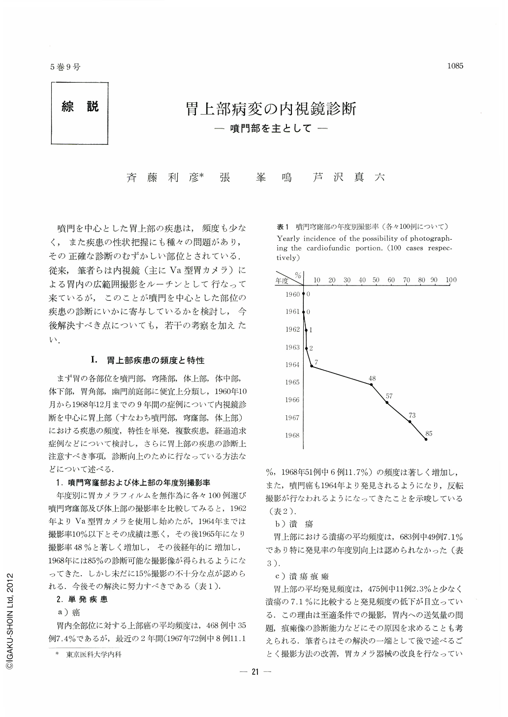
噴門を中心とした胃上部の疾患は,頻度も少なく,また疾患の性状把握にも種々の問題があり,その正確な診断のむずかしい部位とされている.従来,筆者らは内視鏡(主にVa型胃カメラ)による胃内の広範囲撮影をルーチンとして行なって来ているが,このことが噴門を中心とした部位の疾患の診断にいかに寄与しているかを検討し,今後解決すべき点についても,若干の考察を加えたい.
The present paper is a review of lesions in the upper portion of the stomach (the cardia, fundus and upper corpus) encountered in the years 1960~1968. Gastrocamera GT-Va was mainly used for their diagnoses.
Since the examinations are carried out for lesions involving the upper portion of the stomach, a check on the thoroughness of the preliminary gastroscopic examination is required. Any suspicious lesion found by this preliminary examination, even when it is taken by an improper photographic condition such as distant, oblique or partial views, needs a secondary specific detailed examination under a proper photographic technique to establish a correct diagnosis.
The accurate diagnosis of lesions in the upper portion of the stomach needs an abundant experience in differentiating normal pattern of the cardia from abnormal one. Additional information on the fundus (especially diverticulum) can be obtained by removing mucous lake while posture of the patient is changed. The specific photographic technique requires a special gastroscope such as dual tilting gastrocamera type GT-Vw as well as an ordinary gastrocamera GT-Va with combination of “U” and “J” turn methods. Esophagofiberscopy is indicated in lesions located just below the cardia (especially for small cancer and ulcer scar). Observation of these lesions under esophagofiberscoporeven biopsy through the scope is further indicated in some cases. As ulcers in this portion of the stomach always show malignant manifestations even when they are benign in biopsy, close observation and repeated endoscopic examination are strongly advised to distinguish malignant from benign ulcers. Once the diagnosis of benign ulcer is established, follow-up endoscopic observation under the same photographic condition is necessary until its cicatrization is confirmed and a superficial depressive type of early gastric cancer is ruled out.
It is hoped that far greater numbers of lesions in the upper part of the stomach shall be accurately diagnosed and characteristics of early cancer in this portion clarified.
Copyright © 1970, Igaku-Shoin Ltd. All rights reserved.