Japanese
English
- 有料閲覧
- Abstract 文献概要
- 1ページ目 Look Inside
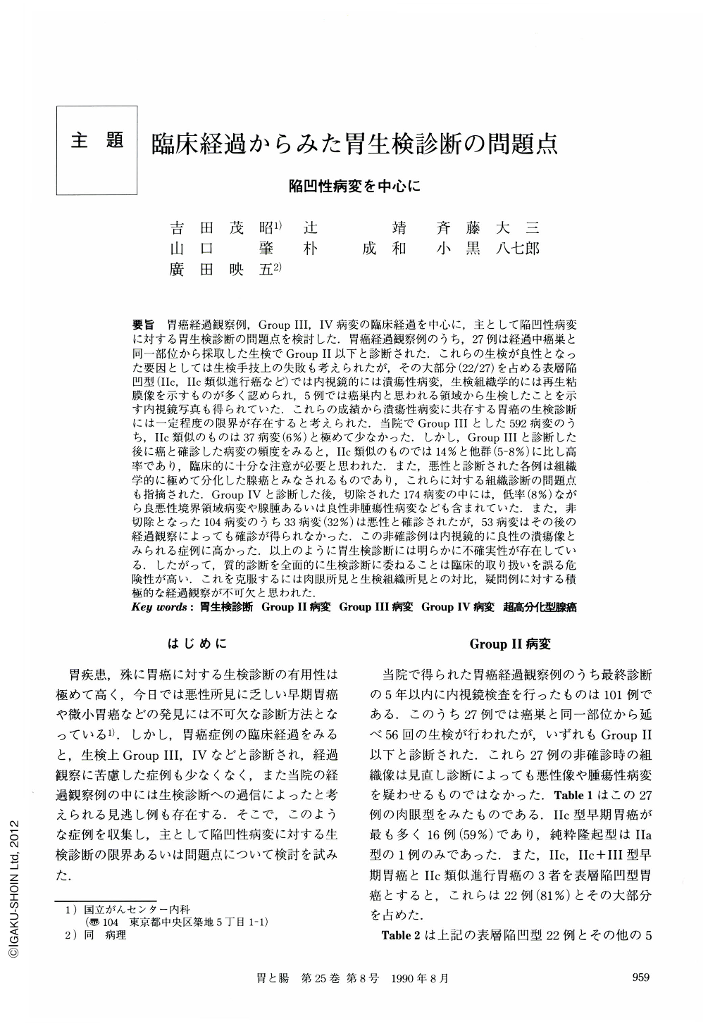
要旨 胃癌経過観察例,GroupⅢ,Ⅳ病変の臨床経過を中心に,主として陥凹性病変に対する胃生検診断の問題点を検討した.胃癌経過観察例のうち,27例は経過中癌巣と同一部位から採取した生検でGroupⅡ以下と診断された.これらの生検が良性となった要因としては生検手技上の失敗も考えられたが,その大部分(22/27)を占める表層陥凹型(Ⅱc,Ⅱc類似進行癌など)では内視鏡的には潰瘍性病変,生検組織学的には再生粘膜像を示すものが多く認められ,5例では癌巣内と思われる領域から生検したことを示す内視鏡写真も得られていた.これらの成績から潰瘍性病変に共存する胃癌の生検診断には一定程度の限界が存在すると考えられた.当院でGroupⅢとした592病変のうち,Ⅱc類似のものは37病変(6%)と極めて少なかった.しかし,GroupⅢと診断した後に癌と確診した病変の頻度をみると,Ⅱc類似のものでは14%と他群(5-8%)に比し高率であり,臨床的に十分な注意が必要と思われた.また,悪性と診断された各例は組織学的に極めて分化した腺癌とみなされるものであり,これらに対する組織診断の問題点も指摘され.GroupⅣと診断した後,切除された174病変の中には,低率(8%)ながら良悪性境界領域病変や腺腫あるいは良性非腫瘍性病変なども含まれていた.また,非切除となった104病変のうち33病変(32%)は悪性と確診されたが,53病変はその後の経過観察によっても確診が得られなかった.この非確診例は内視鏡的に良性の潰瘍像とみられる症例に高かった.以上のように胃生検診断には明らかに不確実性が存在している.したがって,質的診断を全面的に生検診断に委ねることは臨床的取り扱いを誤る危険性が高い.これを克服するには肉眼所見と生検組織所見との対比,疑問例に対する積極的な経過観察が不可欠と思われた.
In order to clarify diagnostic problems of endoscopic biopsy, we examined endoscopic and histopathological features in gastric cancer cases (mostly depressed type). Lesions were classified into benign (Group Ⅰ or Ⅱ), borderline (GroupⅢ) or possible malignant (GroupⅣ) by biopsy throughout the course. In 27 cases of gastric cancer, the lesions detected at the previous examinations during five years were histologically diagnosed as benign. Twenty-two out of these 27 cases were superficially depressed (Ⅱc or Ⅱc-like advanced) type. Most of the lesions in these cases had been considered ulcerative change endoscopically and regenerative mucosa histopathologically. It suggests that most of the previous biopsy specimens were obtained at least from ulcerative lesions, i.e., appropriate biopsy sites. In addition, in five out of these 22 cases, endoscopic pictures of biopsy site showing possible malignant area were available, as shown in Fig. 1. Thus biopsy examination of ulcerative cancer has some limitation in detecting malignancy if the biopsy specimen contains regenerative change by ulceration.
Though only 37 out of 592 lesions diagnosed as GroupⅢ by biopsy were considered as superficially depressed type endoscopically, incidence of malignancy detected by examining follow-up biopsy or resected material was much higher (14%) in these 37 lesions than the rest of the lesions (5-8%). Most of these malignant lesions exhibited histologically only minor structural and cellular atypism which was difficult to differentiate from gastric adenoma. According to these findings, depressed lesions diagnosed as GroupⅢ should be considered possible malignant lesions with only minor histological atypism.
Of the 278 lesions of GroupⅣ, 174 were resected leading to the final diagnosis of borderline or benign lesions in 14 lesions (8%) in spite of the prior biopsy results (possible malignancy). Furthermore, in the remaining 104 unresected lesions malignancy was not confirmed by follow-up biopsy in 53 lesions. And the proportion of malignancy which tended to be elusive was very high among those considered as benign ulcerative lesion endoscopically.
Since these results clearly show limitations of endoscopic biopsy in diagnosing gastric cancer, it is dangerous to totally rely on biopsy results in differentiating benign from malignant lesions. Thus it seemed to be critically important that both endoscopic and histological findings, or follow-up biopsy results should be taken into consideration in the diagnostic process of questionable lesions.

Copyright © 1990, Igaku-Shoin Ltd. All rights reserved.


