Japanese
English
- 有料閲覧
- Abstract 文献概要
- 1ページ目 Look Inside
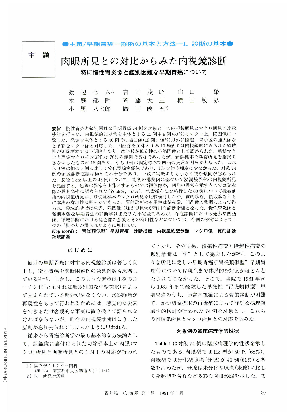
要旨 慢性胃炎と鑑別困難な早期胃癌74例を対象として内視鏡所見とマクロ所見の比較検討を行った.内視鏡的に褪色を主体とする15例中9例(60%)はマクロ上,陥凹像に一致した.発赤を主体とする40例では陥凹像(19例:48%)以外に隆起,胃小区の腫大像など多彩なマクロ像と対応した.凹凸像を主体とする19病変では内視鏡的にみられた領域性が切除標本では不明瞭となり,約半数が孤立性の小陥凹像として認められた.新鮮マクロと固定マクロの対応性は76%の症例で良好であったが,新鮮標本で異常所見を指摘できなかったものが16例あり,うち9例は固定標本で凹凸の異常が明らかとなった.これら9例は他の7例に比して分化型腺癌優位であり,Ⅱbを伴う頻度は少なかった.対象74例の領域診断成績は極めて不十分であり,一般に実際よりも小さく読む傾向が認められた.長径1cm以上の48例について,術後の構築図に基づいて浸潤境界部の内視鏡所見を見直すと,色調の異常を主体とするものでは褪色像が,凹凸の異常を示すものでは発赤像が最も高率に認められた(各59%,67%).色素撒布法を施行した63例について撒布前後の内視鏡所見および切除標本のマクロ所見を比較検討したが,質的診断,領域診断ともに本法の有用性は明らかであった.質的診断の有用性は発赤像,凹凸像の強調によって得られ,領域診断では発赤,陥凹像に加え褪色像が有用な診断指標となった.慢性胃炎像と鑑別困難な早期胃癌の診断学はまだまだ不完全であるが,存在診断における発赤や凹凸像,領域診断における褪色像の意義とその有用性などについては,今回の検討によって1つの手掛かりが得られたように思われた.
Seventy four cases of solitary early gastric cancer whose endoscopic appearance was difficult to differentiate from the appearance of chronic gastritis ("gastritis -like" type) were selected as the subjects of this study. Their preoperative endoscopic appearance and postoperative macroscopic appearance on the resected specimen were compared to evaluate more objectively the diagnostic accuracy of findings of gastritis-like malignancy. Endoscopically the 74 cases were classified into the following three sub-types; flat discolored (D-type; 15cases), flat reddish (R-type; 40 cases) and granular (G-type; 19 cases) ones, as shown in Figures 1, 2 and 3.
As shown in Table 3, in more than half (60%) of the cases of D-type, discoloration corresponded to a depressed area on the freshly resected specimen. Redness in the R-type corresponded to variable macroscopic findings; not only to depression (48%) but also to granularity, elevated components and discoloration. Furthermore, in 30% of this type, no abnormality was detected macroscopiclly in the freshly resected specimens. In the G-type, demarcation of granular lesions observed endoscopically were unclear on the resected material and they were seen as solitary small depressions or elevations of the 16 cases whose cancerous lesion could not be detected on the fresh material macroscopically, 9 were detectable because of their mucosal abnormality on the fixed resected material. In these 9 cases, differentiated type was more dominant histologically, and Ⅱb-like extension was less frequent than in the remaining cases (see Table 5).
Diagnostic accuracy of the lateral extension was very poor in these subjects, and, in general, the size of the cancer was underestimated endoscopically (see Table 6). Retrospective observation of endoscopic findings in the marginal area carried out in 48 cases whose size was more than 1 cm in diameter, revealed that discoloration was the most frequent (59%) finding in D- and R-types, and reddness in G-type (67%) (see Table 7).
Dye spraying endoscopy using 0.1% of Indigocarmine was evidently useful to make both qualitative and quantitative diagnosis of these lesions. It made malignant findings more clear by emphasizing irregularity in reddness, depression and granularity, and made lateral extension clearer by emphasizing depression or discoloration, as shown in Tables 9 to 11.
Endoscopic diagnosis of gastritis-like malignancy is still rather poor. The above results, however, indicated the great usefulness of the combined use of conventional and dye-spraying endoscopy. Also, when we diagnose gastritis-like malignancy, the reddness, or depression observed endoscopicaly can be assessed as indicative of mucosal abnormality, and the margin of discoloration or depression can be regarded as determining the border of lateral invasion.

Copyright © 1991, Igaku-Shoin Ltd. All rights reserved.


