Japanese
English
- 有料閲覧
- Abstract 文献概要
- 1ページ目 Look Inside
Ⅰ.はじめに
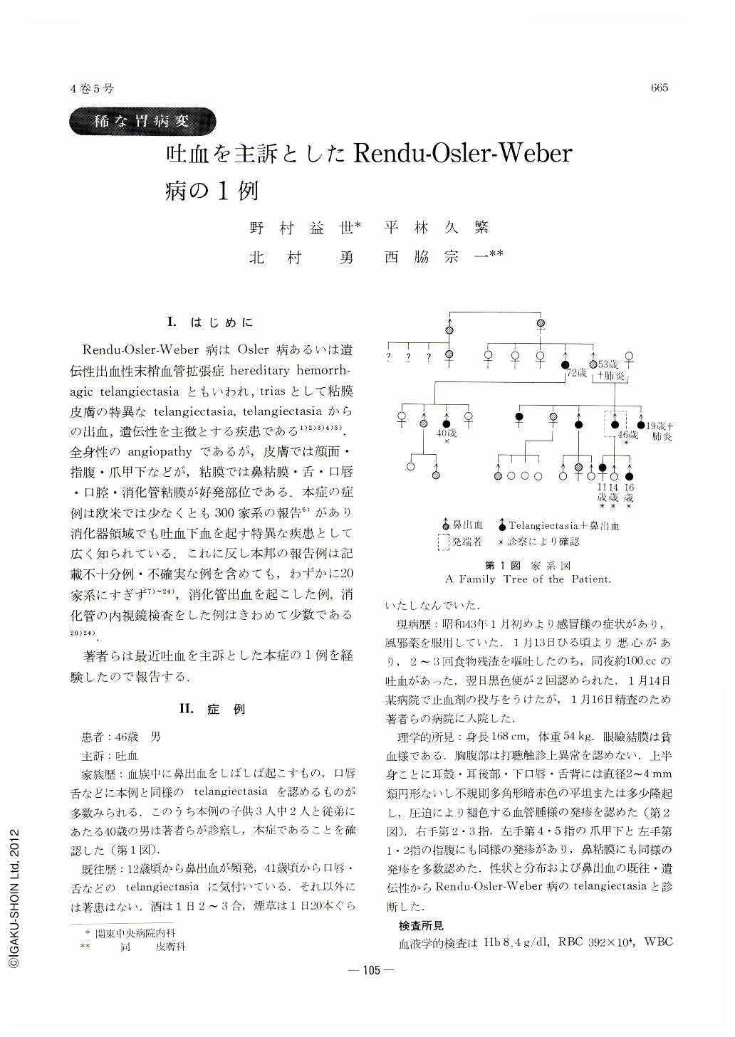
Rendu-osler-Weber病はosler病あるいは遺伝性出血性末梢血管拡張症hereditary hemorrhagic telangiectasiaともいわれ,triasとして粘膜皮膚の特異なtelangiectasia,telangiectasiaからの出血,遺伝性を主徴とする疾患である1)2)3)4)5).全身性のangiopathyであるが,皮膚では顔面・指腹・爪甲下などが,粘膜では鼻粘膜・舌・口唇・口腔・消化管粘膜が好発部位である.本症の症例は欧米では少なくとも300家系の報告6)があり消化器領域でも吐血下血を起す特異な疾患として広く知られている.これに反し本邦の報告例は記載不十分例・不確実な例を含めても,わずかに20家系にすぎず7)~24),消化管出血を起こした例,消化管の内視鏡検査をした例はきわめて少数である20)24).
著者らは最近吐血を主訴とした本症の1例を経験したので報告する.
Rendu-Osler-Weber's disease (Osler's disease or hereditary hemorragic telangiectasia) is commonly known by its trias, namely, specific telangiectasia on the skin and mucous membrane, hemorrahage caused by it and its familial incidence. Predilection sites on the skin are face, the bulbs of fingers and nail bed, and those on the mucous membrane are nasal mucosa, tongue, lip, oral lumen and digestive tract. Preferential sites of other viscera are lung, liver spleen, and so on. In foreign countries this disease is widely known as a peculiar ailment causing hematemesis and melena, found in more than 300 families according to the literature thereon. In this country only about 20 families are known to harbor this disease, and there have been scarcely any report of it associated with gastrointestinal hemorrhage, still less so as to its endoscopy study.
In this paper is presented a case of Rendu-Osler-Weber's disease encountered in a man 46 years of age complaining of vomiting of blood, illustrated with pictures in color of telangiectasia of his stomach. Of three of his children, two siblings and one of their cousins were discovered to have the same variety of telanigiectasia on their nasal membranes, lips, tongues, etc. It is said that bleeding from these sites has often occurred.
Teleangiectasia of the stomach is similar in its gross picture to that of the lip and nose, as it is seen as of rather circular shape, or at times having irregular shape with uneven margin. It is slightly elevated, or sometimes not at all, from the mucosal surface. The gastric mucosa is here and there dotted with these vascular anomalies, having the diameter of from 1 to 10 mm. Though at a first glance they may be mistaken for bleeding spots in the mucosa, the former can be discriminated as the same findings are observed in the same place every time the patient is examined.
In the study of a patient having a bout of hematemesis and/or melena of unknown etiology, telangiectasia should at least be taken into consideration, although it is of rare occurrence.

Copyright © 1969, Igaku-Shoin Ltd. All rights reserved.


