Japanese
English
- 有料閲覧
- Abstract 文献概要
- 1ページ目 Look Inside
- サイト内被引用 Cited by
Ⅰ.はじめに
内視鏡で早期胃癌と診断するためには癌か否かの鑑別と同時に癌の深達度を推定しなければならない.このことは癌細胞の深部浸潤の深さという顕微鏡レベルでの尺度を,内視鏡という肉眼レベルでの粘膜表面の形態的な変化所見から推定しようとするのであるから,もとより難しい問題である.しかし,これが不可能であるならば,癌深達度で規定された1)早期胃癌を内視鏡的に診断することも不可能であるといわねばならない.
従来,早期胃癌の診断学に関する研究は癌か否かの問題がもっぱら議論され癌深達度の推定を系統的に検討した報告は見られず,これを困難とする研究者も少なくないが,本年度福岡での学会以来にわかに活発な議論を呼ぶようになってきた.
著者らは数年来内視鏡学会分類2)3)に亜型分類4)を加えこの問題を検討してきたが4)5)6)7),その後の症例を加えて早期胃癌と誤診しやすい進行癌はどれくらいあるか,それらはどの病型で多いか,両者を鑑別するに役立つ所見としてどういうものがあるか,また内視鏡で”早期”という診断はどの程度可能であるかなどについて検討したので,その成績を以下に述べる.
For endoscopic diagnosis of early gastric cancer, limited to the mucosa or the submucosa in its cancerous involvement, it is necessary to differentiate benign lesion from malignant one as well as to estimate the depths of cancerous invasion.
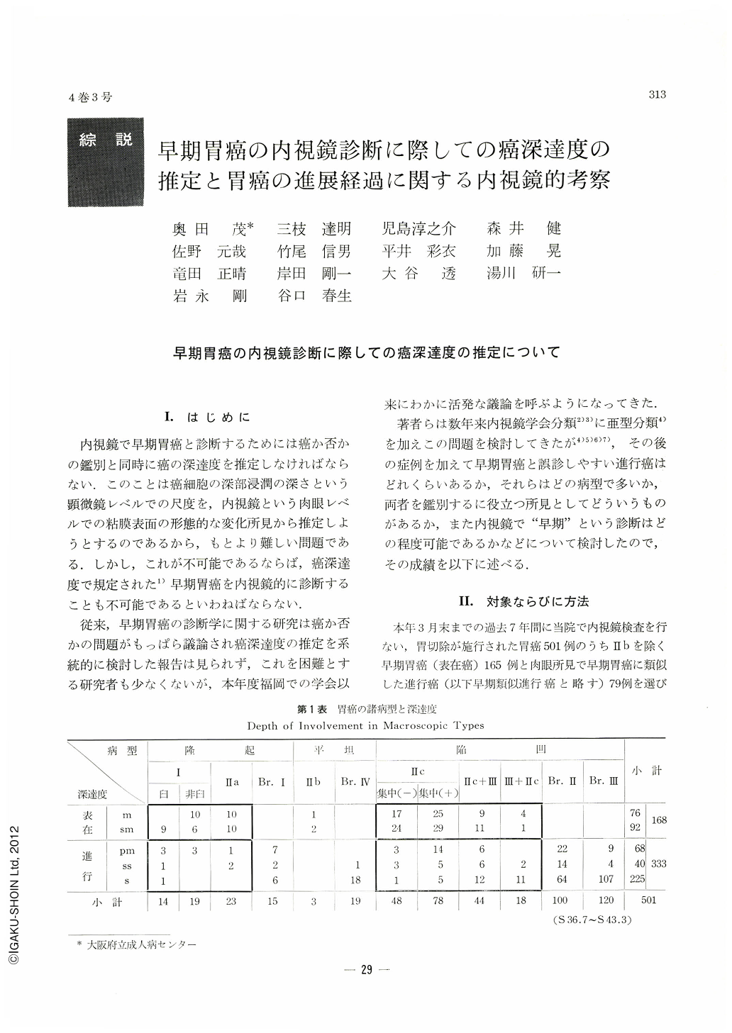
Operations were performod during the period from July, 1961, to March, 1968, on 165 cases of early gastric cancer which were confirmed as such histologically, in addition to 79 cases of advanced cancer whose macroscopic findings simulated those of early gastric cancer. We had subdivided some types of early gastric cancer as reported in previous papers4~11). In each type or subtype, their gross and endoscopical findings were compared with histological data, and criteria have been pursued for the estimation of the cancer involvement. It has also been tried to find endoscopical diagnostic limit for its estimation.
Endoscopic findings clarified so far for estimating the depth of malignant involvement in each type of early gastric cancer are as follows.
Type Ⅰ. In the molar form of Type Ⅰ (molar-shaped protrusion covered with the extension of surrounding mucosa, having broad base and shallow defect at the tip), most cases are proved as submucosal, while that with deeper deficit at the top is mostly advanced. In the non-molar form of Type Ⅰ, cancer lesion, less than 2cm in size, is limited in most instances within the mucosa, and that, larger in size and with broader base, reaches the submucosa or further beyond it.
Type Ⅱa. In this type, lesions with following findings are mostly suhmucosal or advanced, irregular nodularity on the surface, size of more than 4cm in diameter, and coexistence of deep depression.
Type Ⅱc Most of lesions of Type Ⅱc with marked depression are either suhmucosal or advaneed. Type Ⅱc featured hy α finding (remnant of converging folds identifiable inside of Ⅱc lesion with sharp margin) mostly has its invasion limited to the mucosa, but if featured by, β finding (folds disrupted at the margin of Ⅱc lesion; some with enlarged ends), or by γ finding (folds disrupted with confluent ends at the margin of Ⅱc lesion), its invasion reaches the submucosa or beyond it.
Type Ⅱc+Ⅲ. Lesions having a wide crater, more than 2cm in diameter, mostly invade the submucosa or beyond it. Characteristic features of α, β or γ finding at the margin of Ⅱc lesion may be helpful for the estimation of the depth of malignant involvemet.
Type Ⅲ+Ⅱc. Estimation is diificult in this combined type.
In general, erroneous diagnosis was only few in number in cases of various types with elevated lesion and of Type Ⅱc without converging folds, whereas it happened not infrequently in cases of Type Ⅱc with converging folds, Type Ⅱc+Ⅲ and Type Ⅲ+Ⅱc, showing thus a tendency to underestimation in the former and to overestimation in the latter two.
Of all early gastric cancer proved histologically as such, three fourths of them were correctly diagnosed by endoscopic method, while of all early gastric cancer tentatively diagnosed as such by endoscopic study, about four fifths were accurately diagnosed. As for cases of seemingly early but really advanced cancers, about one third of them were mistaken for early ones.
Mode of Development of Early Gastric Cancer Observed by Endoscopy
In this series, 21 early cases as well as 9 advanced cases including 4 of these with an outward appearance of early cancer had one or more previous endoscopic examinations at intervals of six months or more, making it thus possible to do a retrospective study on the mode of development of gastric cancer. The longest interval in these cases was six years and five months. Some of the early cases showed little difference between the findings at preceding examination and those at a later date even after intervals of one to four years, revealing markedly slow development of cancer. Some of the advanced disclosed rapid progression from a faint change to a striking alteration of Borrmann's type in less than three years.
In early gastric cancer, remarkable development was found only in 7 cases (33%), which belonged to the non-molar form of Type Ⅰ, Type ⅡⅡc with converging folds and Type Ⅱc+Ⅲ, while in advanced cancer, it was seen in 8 cases (89%), such as with an appearance of Type Ⅱc, as well as Type Borrmann Ⅱ or Ⅲ.
It was observed by repeated endoscpic examinations that, in general, development in the vertical direction to the level of the mucosa was more conspicuous than that in the horizontal one to it, as exemplified by such findings as more deepening of depressed lesion, increased sharpness of the margin of Ⅱc lesion, transition of the features at the margin of Ⅱc lesion from α to β up to γ findings, formation of an elevated wall, increased irregularity at the bottom of depression or on the surface of elevated lesion, appearance or disappearance of ulceration inside of Ⅱc lesion, etc. By contrast, evidence of superficial spread even in large Ⅱc lesion has never been recognized.
Interrelationship between gastric acid secretion and some histological types of cancer, such as signet ring cell carcinoma and car. simplex, was further referred to in this paper with regard to the process of ulceration inside of Ⅱc lesion.
Copyright © 1969, Igaku-Shoin Ltd. All rights reserved.