Japanese
English
- 有料閲覧
- Abstract 文献概要
- 1ページ目 Look Inside
Ⅰ.はじめに
胃の細胞学的診断法が,その第一歩を踏み出したのは,Papanicolaou1)により試みられた空腹時胃液の細胞診にはじまるといってよいであろう(1946,1947).本邦においては綾部ら(1949)2)が,胃液吸引法による胃癌細胞診に成功し,つづいて,生理的食塩水3)4)による標本採取が試みられたが,当時は尚80%前後或いはそれ以下の陽性率にとどまり,酵素液洗滌法,abrasive ballon法の開拓によって,ようやく85~90%の診断率が報告されるに至った.特に山田(1959)5)のTrypsinによる蛋白融解酵素洗滌法,阪6)7),信田8),山形,石岡ら9)のabrasive balloon法の改良による擦過法が応用されるに至り,胃癌細胞診の臨床的価値が評価されるようになった.しかし早期胃癌例に対する成績は,このような非直視下法では,70~80%の診断率にとどまり,わずかに安藤10)によるX線透視下選択的洗滌法,城所ら11)によるV型胃カメラを改良した洗滌器などにより,多少の進歩がみられたが,早期胃癌の細胞診の飛躍的向上は,春日井ら12)の考按になる直視下洗滌細胞診用ファイバースコープの開発によるものと考える.このような直視下採取法では,非直視下法に比して,早期胃癌,進行胃癌の間にその成績に大差なく,ともに95%前後の診断率があげられ,むしろ早期胃癌のほうが採取し易い症例が多くなり,臨床診断学として,その地位を確立したといってよいであろう12).
本稿の主題である直視下吸引細胞診は,直視下洗滌法,直視下生検法の長所を生かし,欠陥を補う意味で,
1.胃粘膜面の損傷を軽くすること
2.標本の採取,処理,鏡検をより簡易にすること
3.狙撃性を活用すること
等を目的として開発した方法である.
Three methods are used at present in direct vision cytology; the lavage method, the suction method, and contact smears of biopsy tissue. This paper is to discuss the clinical value of these technics with special reference to the suction method.
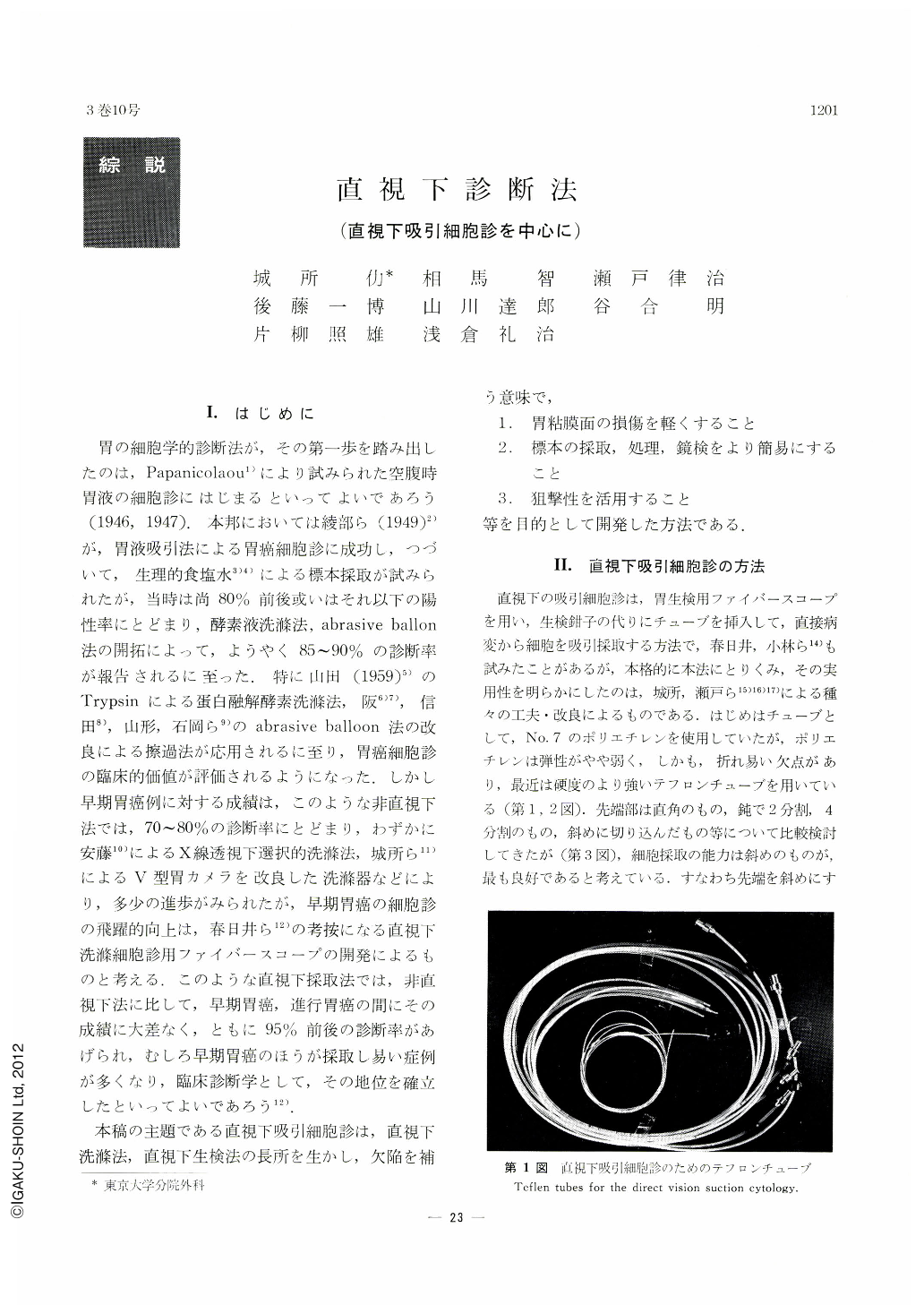
The instrument used for the suction method is the Model B fiberscope (fiberscope for biopsy), with which a thin Teflon tube with a beveled metal tip instead of the biopsy forceps is introduced into the stomach and advanced, under direct vision, to the given target. Repeated negative pressure using a 100 cc syringe is then applied to suck material into the tube. The material is then flushed and smeared on a slide glass.
This method is characterized by the following,
1. Preliminary lavage is not necessary.
2. Abundant fresh cells can be obtained with minimal necrotic debris.
3. Multiple lesions can be examined simply by changing the tube.
4. The damage given to the tissue is least in comparison with the lavage method or biopsy. The method is, therefore, particularly suitable for minute lesions, but it is cliFficult to obtain material from deeper portions.
5. The suction method has given correct diagnosis in 95% of the cases, that is comparable to the other methods. The method thus suffices clinical purposes, but the result will be further improved if these technics are combined acording to the location or morphological characteristics of the lesion to be examined.

Copyright © 1968, Igaku-Shoin Ltd. All rights reserved.


