Japanese
English
- 有料閲覧
- Abstract 文献概要
- 1ページ目 Look Inside
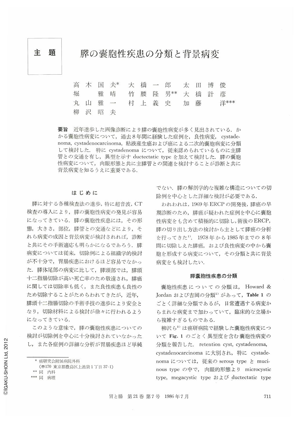
要旨 近年進歩した画像診断により膵の囊胞性病変が多く見出されている.かかる囊胞性病変について,過去8年間に経験した症例を,良性病変,cystadenoma,cystadenocarcinoma,粘液産生癌および癌による二次的囊胞病変に分類して検討した.特にcystadenomaについて,従来認められているものに主膵管との交通を有し,異型を示すductectatic typeを加えて検討した.膵の囊胞性病変について,肉眼形態と共に主膵管との関連を検討することが診断と共に背景病変を知るうえに重要である.
Number of cystic lesion in the pancreas is increasing because of recent development of image diagnostic technique.
Based on 27 cases resected in Cancer Institute Hospital for these 8 years from 1978 to 1985, classification of cystic lesion in the pancreas is proposed as followings, benign lesion, cystadenoma, cystadenocarcinoma, mucous-producing cancer and secondary cystic lesion caused by cancer.
Benign lesion includes 4 cases of pseudocyst and 4 cases of ductogenic cyst.
Cystadenoma involves two classical types, microcystic type and megacystic type, and the original type, ductectatic type which have connection with main pancreatic duct. Ductectatic type located mainly in the uncinate process of the pancreas head and showed characteristic patterns of panereatogram lake as tree root appearance, small diameter from 2 to 4 cm and mucin production.
Cystadenocarcinoma and mucous-producing cancer have something in common with papillary adenocarcinoma and mucin production. Mucous-producing cancer developer in the main pancreatic duct and causes marked dilatation of main pancreatic duct and widening of the orifice of major papilla. Cystadenocarcinoma shows round shape and has no connection with main pancreatic duct. Therefore, clinical discrimination of both became possible.
Cystic change caused by main pancreatic duct obstruction due to cancer is often observed. When cystic change and cancer locate closely, cancerous lesion may be concealed by noticeable cystic change.
Here we stress the morphological analysis of cystic lesion and meticulous diagnosis of presence or absence of cancer and inflammatory change, when cystic lesion of the pancreas is detected. US and CT can reveal morphological appearance of cystic lesion, but hardly demonstrates corelation between main pancreatic duct and cystic lesion.
It is important for the diagnosis of cystic lesion and background to know the connection between cystic lesion and main pancreatic duct as well as pancreatogram close to the lesion.

Copyright © 1986, Igaku-Shoin Ltd. All rights reserved.


