Japanese
English
- 有料閲覧
- Abstract 文献概要
- 1ページ目 Look Inside
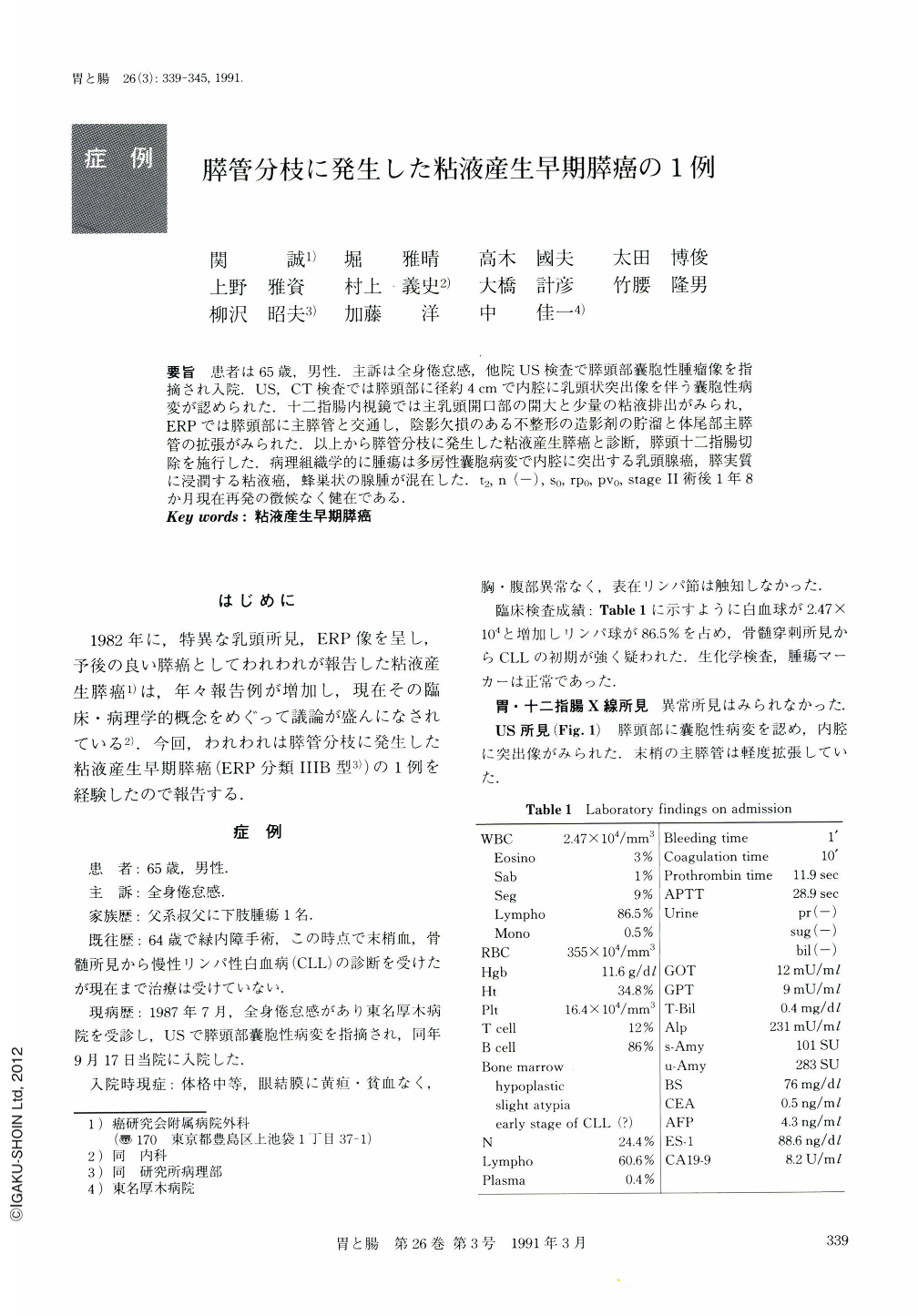
要旨 患者は65歳,男性.主訴は全身倦怠感,他院US検査で膵頭部囊胞性腫瘤像を指摘され入院.US,CT検査では膵頭部に径約4cmで内腔に乳頭状突出像を伴う囊胞性病変が認められた.十二指腸内視鏡では主乳頭開口部の開大と少量の粘液排出がみられ,ERPでは膵頭部に主膵管と交通し,陰影欠損のある不整形の造影剤の貯溜と体尾部主膵管の拡張がみられた.以上から膵管分枝に発生した粘液産生膵癌と診断,膵頭十二指腸切除を施行した.病理組織学的に腫瘍は多房性囊胞病変で内腔に突出する乳頭腺癌,膵実質に浸潤する粘液癌,蜂巣状の腺腫が混在した.t2,n(-),s0,rp0,pv0,stageⅡ術後1年8か月現在再発の徴候なく健在である.
A 65-year-old man complaining of general fatigue was admitted to our hospital for precise examination of a cystic lesion in the head of the pancreas. US and CT revealed a cystic mass with papillary projections. It was about 4cm in diameter (Fig. 1, 2). Endoscopically the orifice of the duodenal major papilla was observed to be opened widely. From it, a small amount of mucin was seen to be excreted. ERP demonstrated irregularly shaped accumulation of contrast medium, and filling defects in the cystic mass (Fig. 3). This lesion communicated with the dilated main pancreatic duct. We finally diagnosed the lesion as mucin-producing pancreatic cancer in a branch of the pancreatic duct. We carried out pancreatoduodenectomy with R2 lymph node dissection. Histologically the tumor was composed of papillary adenocarcinoma projecting into the cystic cavity, mucinous carcinoma infiltrating the surrounding parenchyma of the pancreas, and cystadenoma with slight atypia of the epithelial cells (Fig. 6, 7). Histological diagnosis was mucinous cystadenocarcinoma with adenomatous component, t2, n(-), s0, rp0, pv0, stage Ⅱ. One year and eight months after surgery, the patient is doing well.

Copyright © 1991, Igaku-Shoin Ltd. All rights reserved.


