Japanese
English
- 有料閲覧
- Abstract 文献概要
- 1ページ目 Look Inside
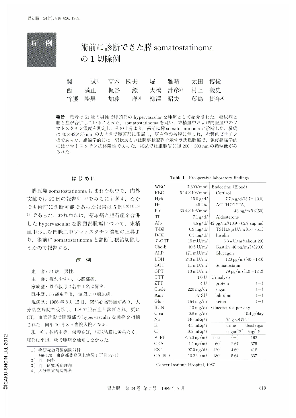
要旨 患者は51歳の男性で膵頭部のhypervascularな腫瘍として紹介された.糖尿病と胆石症が合併していることから,somatostatinomaを疑い,末梢血中および門脈血中のソマトスタチン濃度を測定し,その上昇より,術前に膵somatostatinomaと診断した.腫瘍は40×42×35mmの大きさで膵頭部に限局し,灰白色の被膜に包まれ,赤紫色ゼラチン様であった.組織学的には,索状あるいは腺房状配列を示すラ氏島腫瘍で,免疫組織学的にはソマトスタチン抗体陽性であった.電顕では細胞質に径200~300nmの顆粒像がみられた.
A 51-year-old man was admitted to our hospital with hypervascular tumor of the pancreas head. We suspected pancreatic Somatostatinoma, because his complaint was accompained by diabetes mellitus and cholecystolithiasis.
Then we examined the serum somatostain concentration of the peripheral and portal vein (Fig. 4). Consequently, we were able to diagnose the case as somatostatinoma preoperatively, due to the higher serum somatostatin level than is found normally.
The resected material showed that the tumor was 40×42×35 mm in size, localized at the pancreas head, covered by a grayish white capsule, and having purple and gelatinous parenchyma (Figs. 5 and 7).
Histologically, it was an islet-cell tumor with trabecular and rosette pattern and, immunohistologically, it was stained by anti-somatostatin body (Figs. 8 and 9). Electron microscopy showed fine granules about 200 to 350 nm in diameter in the cytoplasm (Fig. 10) .

Copyright © 1989, Igaku-Shoin Ltd. All rights reserved.


