Japanese
English
- 有料閲覧
- Abstract 文献概要
- 1ページ目 Look Inside
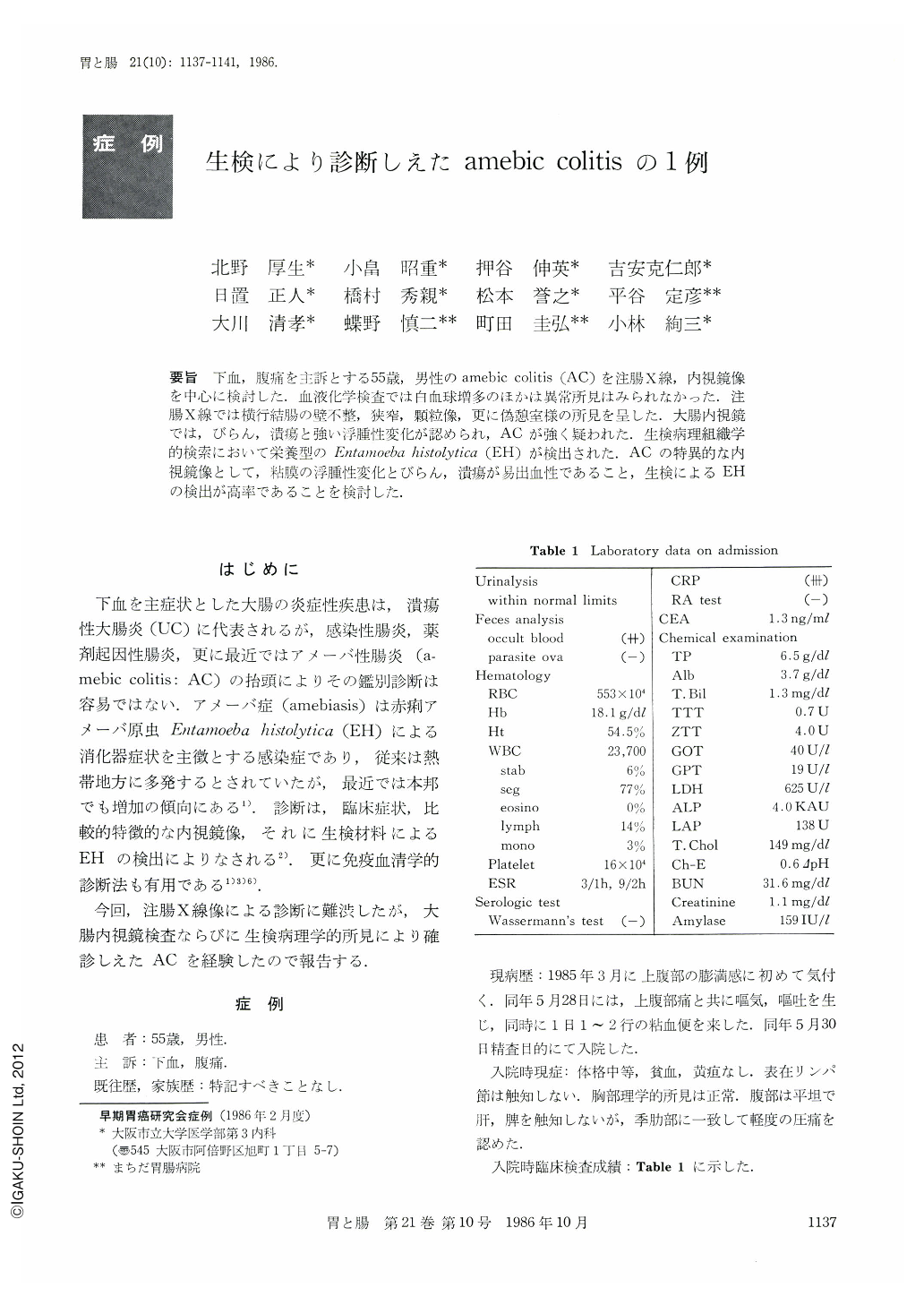
要旨 下血,腹痛を主訴とする55歳,男性のamebic colitis(AC)を注腸X線,内視鏡像を中心に検討した.血液化学検査では白血球増多のほかは異常所見はみられなかった.注腸X線では横行結腸の壁不整,狭窄,顆粒像,更に偽憩室様の所見を呈した.大腸内視鏡では,びらん,潰瘍と強い浮腫性変化が認められ,ACが強く疑われた.生検病理組織学的検索において栄養型のEntamoeba histolytica(EH)が検出された.ACの特異的な内視鏡像として,粘膜の浮腫性変化とびらん,潰瘍が易出血性であること,生検によるEHの検出が高率であることを検討した.
We reported a 55-year-old man who presented with bloody stool and abdominal pain and was finally diagnosed as having amebic colitis. Barium enema showed narrowing and deformity in the transverse colon. Colonoscopy revealed edematous and friable mucosa, providing biopsy specimen which contained Entamoeba histolytica pathognomonic of amebic colitis. Thus, colonoscopic biopsy is one of the useful diagnostic procedures in detecting Entamoeba histolytica as is stool examination.

Copyright © 1986, Igaku-Shoin Ltd. All rights reserved.


