Japanese
English
- 有料閲覧
- Abstract 文献概要
- 1ページ目 Look Inside
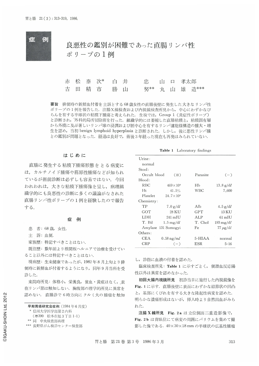
要旨 排便時の新鮮血付着を主訴とする68歳女性の直腸後壁に発生した大きなリンパ性ポリープの1例を報告した.注腸X線検査および内視鏡検査所見から,中心にわずかなびらんを有する半球状の粘膜下腫瘍と考えられた.生検で,Group 1(炎症性ポリープ)と診断され,外科的局所切除術を行った,組織学的には萎縮した直腸粘膜と,粘膜固有層から外膜に及ぶ著しいリンパ球の浸潤および胚中心を有するリンパ濾胞様構造の腫大・増生を認め,当初benign lymphoid hyperplasiaと診断された.しかし,後に悪性リンパ腫との鑑別が問題となった.経過は良好で,術後3年経った現在も再発はみられていない.
A 68 year-old woman was admitted to Shinshu University Hospital complaining of bloody stool in August 1982. Digital examination revealed a walnutsized tumor on the posterior wall of the rectum. Barium enema study showed a hemispherical, partially lobulated, protruded lesion with a smooth surface measuring approximately 40×30mm in width and 18mm in height. Endoscopically the tumor was covered by apparently nomal rectal mucosa with a shallow erosion in its vertex. These findings led to a clinical diagnosis of submucosal tumor in the rectum. Biopsy specimens revealed that the mucosa was slightly atrophic, but the lamina propria showed a marked infiltration by lymphocytic cells. The pathological diagnosis was “benign inflammatory polyp”. To obtain materials sufficient for making a conclusive pathological diagnosis, endoscopic partial resection was carried out by employing high-frequency waves. The histological findings, however, were essentially similar to those of the former specimens. A local excision was performed in January 1983.
The tumor had a broad base and artificial ulceration. Histologically, it was covered by atrophic rectal mucosa except for the partial ulceration. Lymphocytes and plasma cells infiltrated estensively to the lamina propria and submucosa. Furthermore, lymphocytic cells massively infiltrated to the muscularis propria as well as to the adventitial region. The lymph follicle was sparsely presented. The infiltrating lymphocytic cells were relatively mature and mitotic figures were absent except in a geminal center.
Largeness and massive infiltration of the muscularis propria were controversial points in diagnosing whether, in this case, the tumor was a benign lymphoid polyp or malignant lymphoma. There were various discussions about the distinction between benign lymhoid polyp and malignantl ymphoma.
The patient is still alive and well three years after surgery.

Copyright © 1986, Igaku-Shoin Ltd. All rights reserved.


