Japanese
English
- 有料閲覧
- Abstract 文献概要
- 1ページ目 Look Inside
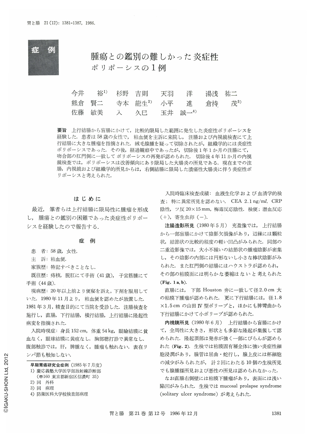
要旨 上行結腸から盲腸にかけて,比較的限局した範囲に発生した炎症性ポリポーシスを経験した.患者は58歳の女性で,粘血便を主訴に来院し,注腸および内視鏡検査にて上行結腸に大きな腫瘤を指摘された.絨毛腺腫を疑って切除されたが,組織学的には炎症性ポリポーシスであった.その後,経過観察中であったが,切除後1年1か月の注腸にて,吻合部の肛門側に一致してポリポーシスの再発が認められた.切除後4年11か月の内視鏡検査では,ポリポーシスは改善傾向にあり限局した大腸炎の所見である.現在までの注腸,内視鏡および組織学的所見からは,右側結腸に限局した潰瘍性大腸炎に伴う炎症性ポリポーシスと考えられた.
We experienced a case with localized giant pseudopolyposis of the colon simulating a neoplastic change.
A 58-year-old Japanese woman with a history of constipation for nearly 20 years was admitted to the Keio University Hospital because of bloody mucus in the stool in 1980. Barium enema and colonoscopy revealed tumor-like polypoid lesion in the ascending colon (Figs. 1 and 2). Ileocecal resection was carried out. The resected specimen showed a giant cluster of nodular, villose, and filiform lesion measuring 8 cm in length and 11 cm in diameter (Fig. 3). Histologic examination revealed the localized area of inflammatory pseudopolyposis without neoplastic change. The mucosa and submucosa were densely infiltrated by inflammatory cells containing foreign body giant cells. But there was no sign of extension of these inflammatory change beyond the muscle layer (Fig. 4).
In 1981, barium enema and colonoscopy demonstrated a recurrence of polyposis in the anal side of the anastomosis (Figs. 5 and 6). The patient was asymptomatic, though. Mucosal biopsies showed inflammatory change. In 1985, after following up for 4 years and 11 months, colonoscopic study showed the features of the localized colitis with only a few polyp formation (Fig. 8).
We concluded that this localized pseudopolyposis of the ascending colon and cecum may be due to the right-sided ulcerative colitis.

Copyright © 1986, Igaku-Shoin Ltd. All rights reserved.


