Japanese
English
- 有料閲覧
- Abstract 文献概要
- 1ページ目 Look Inside
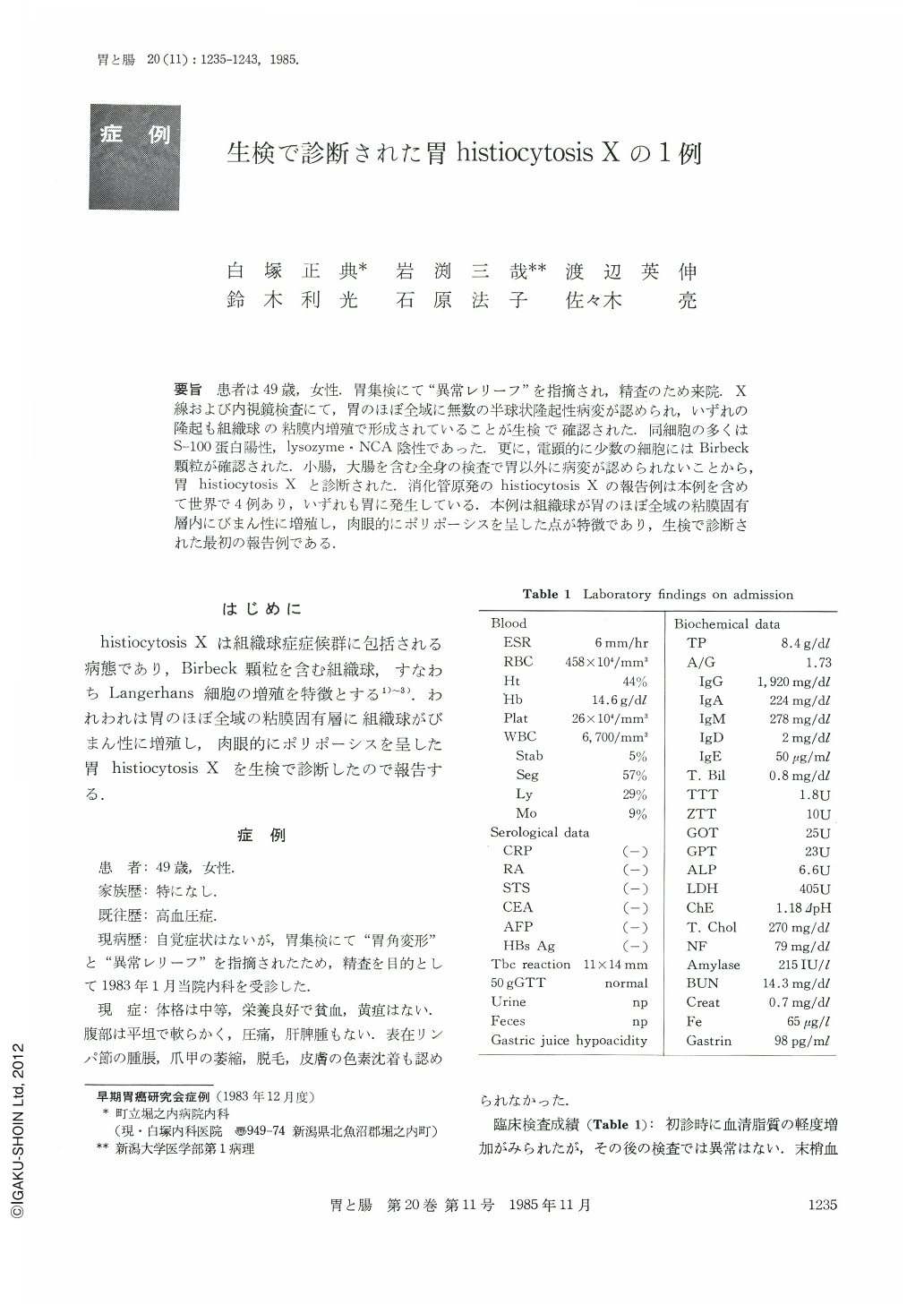
要旨 患者は49歳女性.胃集検にて“異常レリーフ”を指摘され,精査のため来院.X線および内視鏡検査にて,胃のほぼ全域に無数の半球状隆起性病変が認められ,いずれの隆起も組織球の粘膜内増殖で形成されていることが生検で確認された.同細胞の多くはS-100蛋白陽性,lysozyme・NCA陰性であった.更に,電顕的に少数の細胞にはBirbeck顆粒が確認された.小腸,大腸を含む全身の検査で胃以外に病変が認められないことから,胃histiocytosis Xと診断された.消化管原発のhistiocytosis Xの報告例は本例を含めて世界で4例あり,いずれも胃に発生している.本例は組織球が胃のほぼ全域の粘膜固有層内にびまん性に増殖し,肉眼的にポリポーシスを呈した点が特徴であり,生検で診断された最初の報告例である.
A 49 year-old Japanese woman, with no particular complaints, was referred to our hospital on January 1983. Physical examination and study of the blood, urine and feces showed no remarkable changes. X-ray and endoscopic examination revealed innumerable elevated lesions throughout the stomach. The biopsy specimens proved the elevated lesions to be formed by diffuse proliferation of histiocytoid cells in the lamina propria mucosae. They had abundant, slightly eosinophilic cytoplasm and eccentric, ovoid to irregularly indented, vesicular nuclei with small nucleoli. Many histiocytoid cells contained S-100 protein diffusely in their cytoplasm. Lysozyme and α1 alantitrypsin were negative in most of them except for a few cells showing faintly positive reaction. No histiocytoid cells showed CEA or nonspecific antigen cross-reacting with carcinoembryonic antigen. Ultrastructurally some of them contained a single or clusters of Birbeck granules in their cytoplasm mainly at the periphery. Systemic examination by x-ray, endoscopy and computed tomography revealed no abnormality outside the stomach. Therefore the present case was diagnosed as primary histiocytosis X of the stomach.
The patient has been followed up without therapy. Re-examinations by x-ray, endoscopy and biopsy showed no significant changes macroscopically and microscopically. In June 1984, 18 months after the diagnosis, she has continued to be well without complaints.
Only four cases, including ours, are reported as primary histiocytosis X of the stomach. This is the fiirst case that extended throughout the stomach in fashion of polyposis and that was diagnosed with biopsy.

Copyright © 1985, Igaku-Shoin Ltd. All rights reserved.


