Japanese
English
- 有料閲覧
- Abstract 文献概要
- 1ページ目 Look Inside
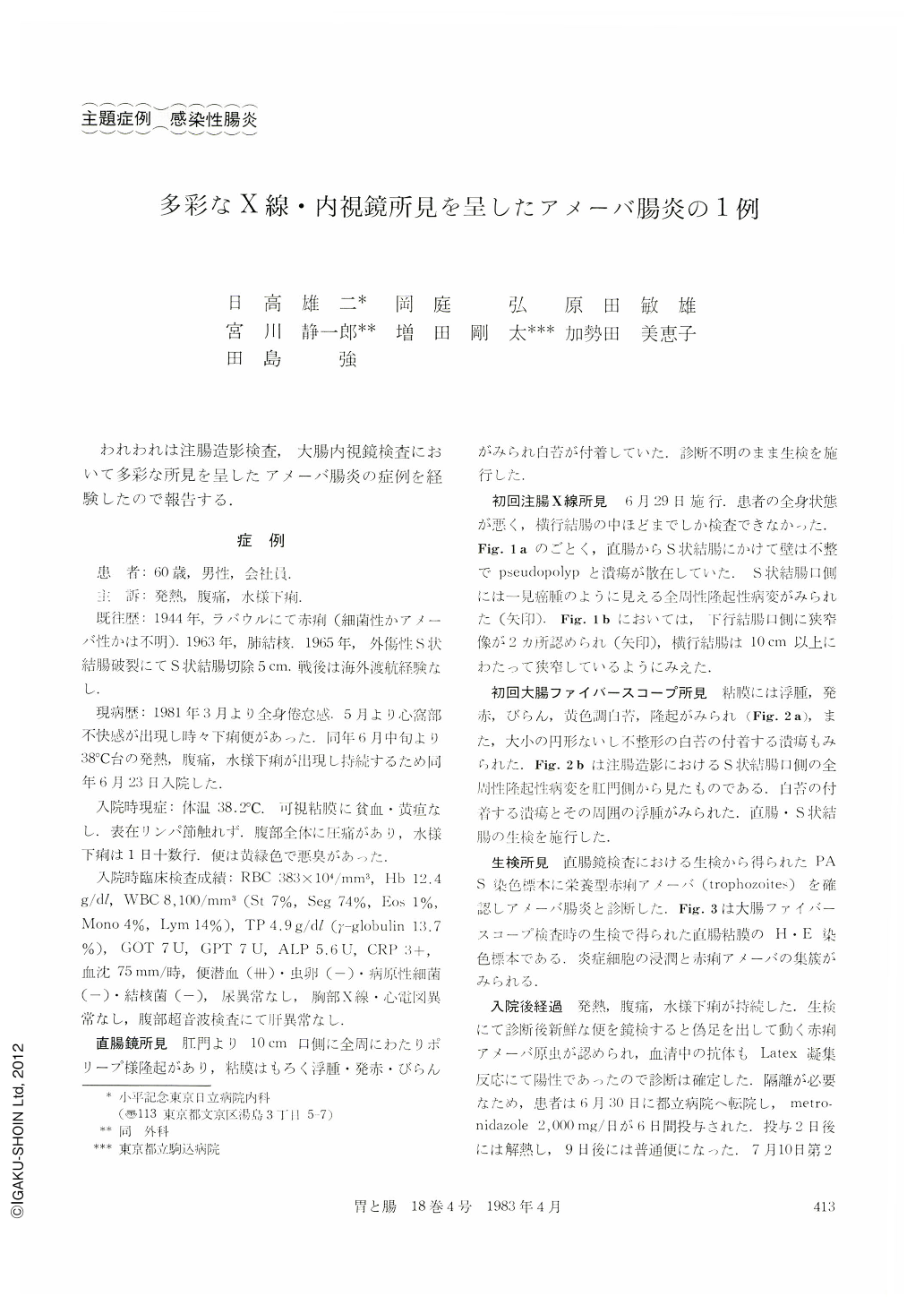
われわれは注腸造影検査,大腸内視鏡検査において多彩な所見を呈したアメーバ腸炎の症例を経験したので報告する.
A 60 year-old man was admitted to our hospital on July 23, 1981, with complaints of persistent fever, abdominal pain and watery diarrhea for five days. He had histories of dysentery in Polynesia in 1944 and pulmonary tuberculosis in 1963, and had a partial sigmoidectomy under the diagnosis of traumatic rupture of sigmoid colon.
Laboratory studies included a red blood cell count of 383×104/mm3, a hemoglobin of 12.4g/dl, a leukocyte count of 8,100/mm3 with a differential count of polymorphonuclear cell 81%, lymphocyte 14%, monocyte 4%, eosinophyl 1%, an erythrocyte sedimentation rate of 75 mm/hr. Chemistry screening was normal except for a total protein of 4.9g/dl.
Proctoscopy revealed inflamed, ulcerated and friable rectal mucosa and the biopsy was done.
Barium contrast study showed multiple pseudopolyps and ulcerations with whitish bases in the rectum and sigmoid colon, and two constricted regions of the descending colon.
Colonofiberscopy demonstrated that the mucosa was edematous and friable, bled slightly and erosive, and there were several white-based ulcerations, 5~10 mm in diameter in the rectum and sigmoid colon.
We diagnosed the patient as having acute amebic colitis because of histologic examination with necrotic debris containing Entamoeba histolytica trophozoites and a positive serologic test. After making the diagnosis, we microscopically observed a mobile ameba in fresh stool.
Following a normal liver ultrasonography, he was transferred to the metropolitan hospital, where treatment with metronidazole 2 g per day for 6 days resulted in complete resolution of his symptoms. Radiologic examination and colonofiberscopy after treatment revealed the disappearance of ulcers.
Because the clinical, laboratory, radiologic and endoscopic features of amebic dysentery are nonspecific, the demonstration of amebic trophozoites in fresh stool is the most direct method of diagnosis. Colonic biopsy is useful even when multiple previous stool examinations are negative. A serologic test for ameba is also helpful. A positive test raises the possibility that the patient has amebiasis when stool examination and biopsies are negative.

Copyright © 1983, Igaku-Shoin Ltd. All rights reserved.


