Japanese
English
- 有料閲覧
- Abstract 文献概要
- 1ページ目 Look Inside
最近の胃診断学の進歩によって,いわゆるびらん性胃炎がにわかに注目されている.その肉眼像は特異であり,タコイボ型,ポリープ型,蛇行型,棍棒型などがあげられている1).筆者らは最近,いわゆるタコイボ型びらん性胃炎のX線ならびに内視鏡像ときわめて酷似した若年者胃転移癌の1例を経験したので報告する.
症例
患 者:岡○美○子 22歳 女
主 訴:心窩部鈍痛.
家族歴;特記すべきことなし.
既往歴:昭和44年8月(当時20歳),当科で横行結腸癌の診断にて結腸右半切除を施行,6×4.5cm大のBorrmannⅡ型の潰瘍形成癌で,組織学的には全層を侵す粘液細胞結節癌(mucocellulonodular carcinoma)を示した(Fig. 8).腫瘍細胞のリンパ管侵入像が散見され,また稀ながら静脈内侵入像も認めた(Fig. 9).領域リンパ節25個のうち,5個のものに同様組織像からなる転移を認めた.Dukes Cの癌であり,断端部への癌細胞の波及は認めなかった.術後化学療法を施行し,46病日に退院し,以後経過良好であった.なお,手術後の胃X線検査では全く異常を認められなかった.
A case of metastatic carcinoma of the stomach is described which on the x-ray and endoscopic findings showed a very close resemblance to so-called gastritis erosiva or verrucosa, one of recent topics in the modern gastric diagnostics.
The patient is a 22-year-old female who underwent right colectomy about two years ago because of primary carcinoma of the transverse colon. Histologically, the tumor consisted of mucocellular nodular tissue which invaded the entire wall with regional lymph node metastases. The stomach was free from any abnormality then.
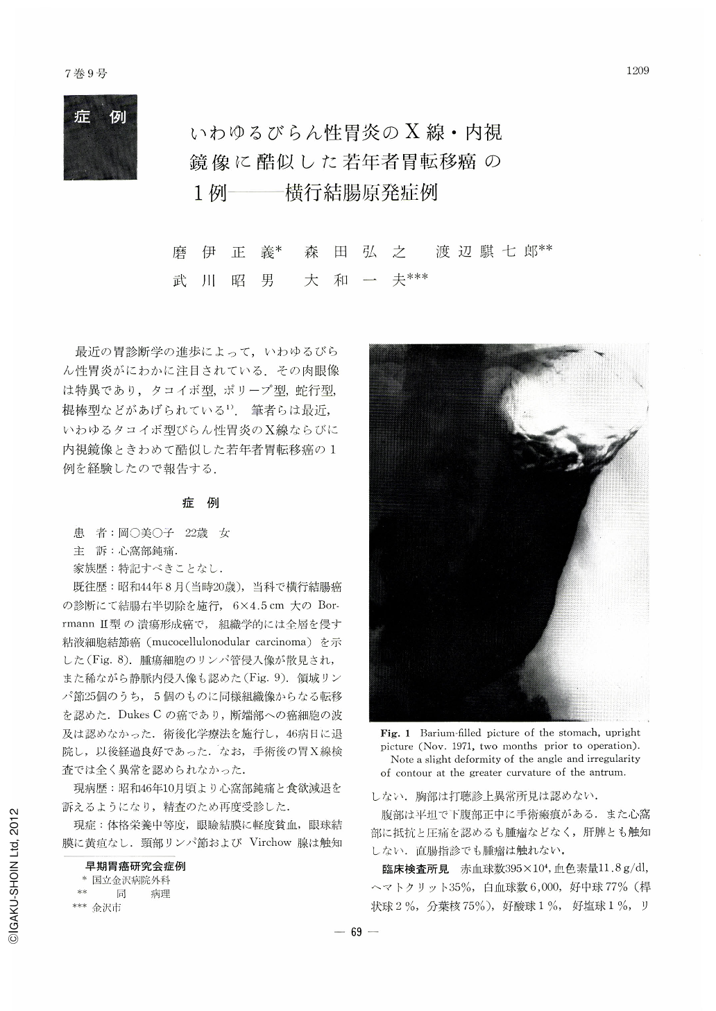
The postoperative course was uneventful and the patient remained well until two years and three months later when she revisited the hospital complaining of dull epigastric pain. X-ray and endoscopy of the stomach revealed a lesion, 4.4×4.5 cm, on the anterior wall of the antrum in addition to multiple small nodules spreading almost all over the stomach. Both lesions were centrally depressed, showing characteristic pictures of so-called gastritis verrucosa. The patient was followed up under that tentative diagnosis.
Re-examination of the stomach about two months later disclosed multiple small nodules increased in number as well as in size. Endoscopic biopsy taken from both lesions revealed signet-ring cell carcinoma. Laparotomy was accordingly performed. Although the serosal surface of the stomach was smooth, many metastatic lymph nodes were palpable in the omentum and mesentery. One of the small nodules in the stomach resected for pathological examination disclosed signet ring cell carcinoma involving both the mucosa and submucosa. The tumor was sharply demarcated against the adjacent intact mucosa with no signs of cellular reaction or glandular metaplasia. Alcian blue-PAS stained sections of the stomach nodule showed the same staining of mucinous carcinoma cells as those of the primary tumor of the colon, so that the former was diagnosed as metastasis from the latter. The patient, treated with chemotherapy, is now under observation for three months after laparotomy.
Because of no documented case of this kind reported so far in the literature, emphasis is laid on a rsemblance of this special case to gastritis verrucosa in the x-ray and endoscopic findings. This fact may contribute to the differential diagnosis of similar lesions.

Copyright © 1972, Igaku-Shoin Ltd. All rights reserved.


