Japanese
English
- 有料閲覧
- Abstract 文献概要
- 1ページ目 Look Inside
かつて,外国人が胃潰瘍の質的診断に非常に興味をもっているという話を聞いて,奇異な感じをもったことがある,当時,私どもの第一の関心事は,より小さな,より微細な早期癌の診断ということであったから,潰瘍辺縁にある癌はすでに小さな病変ではなかったわけである.それに,Ⅲ+Ⅱcの症例が意外に少なかった.
ところが,昨年,本誌でも,Ⅱb型早期胃癌についで,Ⅲ型早期胃癌が特集され,ついで悪性サイクルが特集された.主題も症例も座談会の記事も興味深かった.そして,最近,国立がんセンターの市川平三郎先生から,胃潰瘍の良性・悪性の鑑別診断についての,外国人とわれわれとの間の差を解説してもらって,なるほどと感じ入った.そのような立場からの特集は本誌にもないようである.
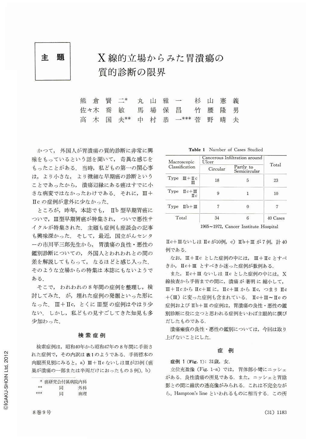
A radiological study was made on the differential diagnosis of malignant and benign ulcers of the stomach. Forty cases of excavated early gastric cancer were used as the material of this study. These cases which had been operated upon in the period of 8 years from 1965 to 1972, included 23 cases of Type III+IIc or III, 10 cases of IIc+III and 7 cases of IIb+III. The following result was obtained.
1) On barium-filled images, niche was observed in only 16 of 40 cases and at least, 10 cases of them were diagnosed as benign ulcer. In the remaining 6 cases, the niches were not characteristic as to make diagnosis of early cancer. The differential diagnosis of malignant and benign ulcers could not be established with the barium-filled image alone.
2-a) Double contrast and compression study was performed in 22 of 23 cases of Type III+IIc and Type III. In 17 cases of them, niche and IIc could not be distinguished radiologically in the whole lesion. In 14 of 17 cases, the whole shape of the lesion was irregular, including ulcer and IIc. In 3 of 17 cases, irregularity of their sharp was in the intermediate degree. In 3 cases, cancer was discovered at the small part of the ulcer margin. In 1 of them, the niche had an irregular outline and in 2 of them, the niche had irregularity in the intermediate degree. Niche and IIc could be distinguished radiologically in 4 cases. In 1 of them, both niche and IIc had an irregular outline. In 2 of them, the lesion as a whole had irregular outline with smoothly outlined niche. In the remaining one case, IIc was discovered at the small part of the ulcer margin.
The diagnosis of Type III+IIc could not be established in 2 cases. In one case, the examination was incomplete due to severe symptoms of the patient, and in the other case the lesion was one of the multiple cancers that escaped detection before surgery.
The most characteristic radiological finding of Type III+IIc and Type III is an irregularly outlined shadow that is best demonstrated by compression method. The double contrast examination is second effective method. When a lesion is big, it may be sometimes diagnosed as an advanced cancer. When a lesion is small, sometimes difficulty arises in differentiating it from Type IIc.
2-b) In all 10 cases of Type IIc+III, niche and IIc was distinguished. Nine cases of them had irregular niche and in one of them irregularity of the niche was in the intermediate degree. The double contrast method is more effective than compression method in order to demonstrate Type IIc+III, because size of Type IIc+III is relatively bigger than Type III+IIc.
2-c) In 7 cases of Type IIb+III, 2 cases had irregular niche; 2 cases had irregular niche in the intermediate degree and 3 cases had smooth niche. Radiological demonstration of a part of IIb was successful in 6 cases.
In conclusion, the most useful radiological sign for the differential diagnosis of gastric ulcer is an irregular outline of its shadow. Once a ulcer is discovered, demonstration of wider area including not only the ulcer itself but also its surrounding mucosa is required. Then, its outline and small IIc at its margin should be demonstrated clearly by compression method. Radiological differential diagnosis of malignant and benign ulcers may be established, if these two method are to be done effectively.
Copyright © 1973, Igaku-Shoin Ltd. All rights reserved.