Japanese
English
- 有料閲覧
- Abstract 文献概要
- 1ページ目 Look Inside
- サイト内被引用 Cited by
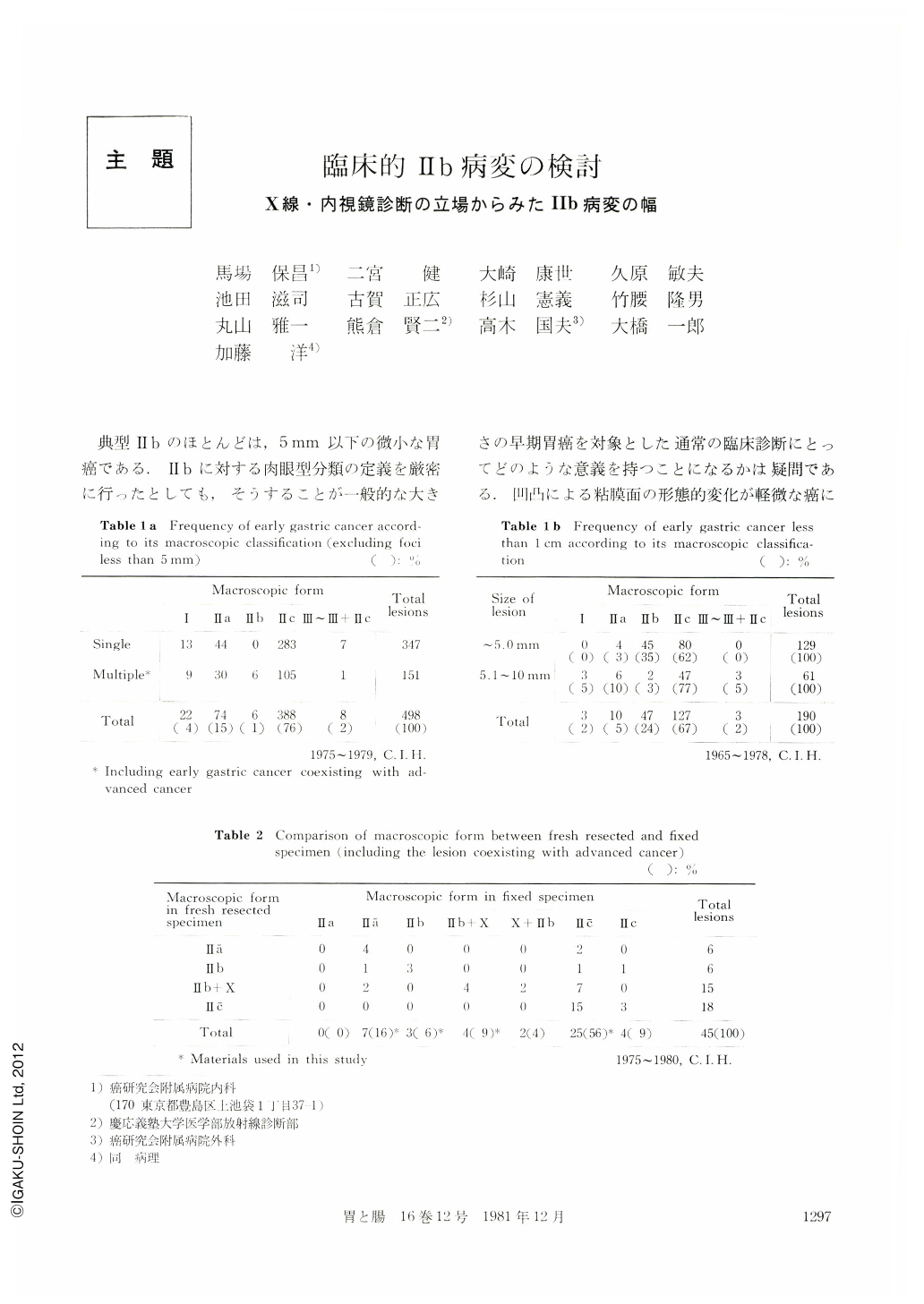
典型Ⅱbのほとんどは,5mm以下の微小な胃癌である.Ⅱbに対する肉眼型分類の定義を厳密に行ったとしても,そうすることが一般的な大きさの早期胃癌を対象とした通常の臨床診断にとってどのような意義を持つことになるかは疑問である.凹凸による粘膜面の形態的変化が軽微な癌に対するX線・内視鏡的な診断限界を追求することを目的とした場合には,Ⅱbの定義を厳しくしておくことも確かに必要である.ところが,そうすると冒頭に述べたようにⅡbのほとんどは5mm以下の微小な胃癌しか存在しないことになり,通常のX線・内視鏡診断にとっては極めてまれな病変でしかなく実際的ではないことになる(Table1,2を参照).一方,一般的な大きさの胃癌の中には,周囲粘膜に対して肉眼的に多少の高低の差があっても,X線・内視鏡的には存在診断あるいは質的診断が困難な癌も少なくはない.このような病変は,ⅡbをX線・内視鏡的に診断が困難である癌と解釈することによって,臨床診断にとってのⅡbということになる.こういったことからは,Hbを臨床的な立場から眺め,X線・内視鏡診断にとって診断が難しい癌とみなして検討することも有意義であるように思われる.
以上のような観点から,本稿ではⅡbを臨床診断の立場から“肉眼的には周囲粘膜に対して軽度な高低の差が認められても,X線・内視鏡的に診断が困難である癌”と定義し,この臨床的なⅡbにはどのような病変が対象となりうるか,そしてそれらはどのようなX線・内視鏡所見ならびに組織学的所見を示しているかについて検討し,臨床的なⅡbに対しての診断の指標を求めてみたい.
Typical type Ⅱb lesions have been incidentally detected in the course of operating on the stomach for some other reason and have a been smaller than 0.5cm. They do not become a target of the clinical diagnosis. In this paper a concept of Ⅱb-like lesion was employed in order to include clinically detected lesions which macroscopically revealed a subtle difference in elevation or depression from the normal surrounding mucosa on the fixed specimen and consequently without clear distinction of their border. In other words, Ⅱb-like lesion means that which is difficult to diagnose radiographically and endoscopically. The radiographic and endoscopic findings of the typical Ⅱb and Ⅱb-like lesion were studied in correlation with their macroscopic and histologic findings.
Materials
Thirty-nine lesions were selected as the typical Ⅱb or Ⅱb-like lesions in a series of 593 histologically proved lesions of gastric cancer which had been operated on from 1975 to 1980 at the Cancer Institute Hospital, excluding the foci less than 5mm.
They were subclassified into 4 varieties; namely Ⅱa (faintly elevated from the normal surrounding mucosa but not so much as Ⅱa) 7 lesions (18%), Ⅱb (typical forms 3 lesions (8%), Ⅱc (faintly depressed but not so much as Ⅱc) 25 lesions (64%) and Ⅱb+Ⅹ (coexisting form, the diameter of Ⅱb should be at least three times as large as that of Ⅹ which is smaller than 2cm) 4 lesions (10%).
Results and discussion
1. Radiographic and endoscopic detection in the initial examination
Fourteen out of 23 lesions which were not associated with ulcerative change were missed, comprising 61% (14/23). On the other hand, all lesions which were associated with ulcerative change were detected. Based on the above mentioned facts, Ⅱa lesion with faint elevation and Ⅱc lesion with faint depression which are not associated with ulcerative change may be defined as Ⅱb lesion in a clinical sense.
2. Determination of malignancy by retrospective review of preoperative radiographic and endoscopic findings
There was no statistically significant relation between presence of ulcerative change and determination of malignancy. There was a statistically significant relation between determination of malignancy and grade of microerosions. Based on above mentioned facts, Ⅱa lesion with faint depression which are accompanied with slight microerosions may be defined as Ⅱb lesion in a clinical sense, irrespective of presence or absence of ulcerative change.
3. Radiographic and endoscopic findings of Ⅱb-like lesion
Although Ⅱb-like lesions revealed only slight mucosal abnormalities in the macroscopic findings, microerosions, and its associated inflammatory and regenerative change were histologically found on the surface of them. And those histologic characteristics were partially reflected in the radiographic and endoscopic finding. In the radiographic findings of the lesions in which determination of malignancy was difficult difference in elevation and depression was delineated more prominently as it was observed in the macroscopic findings. Especially, tiny deep flecks in a faint collection of the barium and partial disappearance or disarrangement of areae gastricae pattern was characteristically visualised.
In the endoscopic findings of the lesions in which determination of malignancy was difficult there was no clear distinction between the abnormal mucosa and the normal mucosa. They only showed nonspecific findings such as punctate redness, punctate redness in a discoloration, and obscure or disturbed pattern of the capillary vessels.
Conclusion
In the radiographic findings of Ⅱb-ike lesion difference in elevation and depression was delineated more prominently as it was observed in the macroscopic findings, on the ground that optimal barium coating was achieved by repeated postural changes. In the endoscopic findings the dye-contrast method was eflective for making the mucosal abnormalities clearer. However, the normal mucosa could not often be distinguished from the lesions. Therefore for the better diagnosis of Ⅱb-like lesion further radiographic and endoscopic investigation should be directed to analysis of the findings which were caused by atrophic, erosive and hyperplastic changes in the noncancerous mucosa.
Copyright © 1981, Igaku-Shoin Ltd. All rights reserved.

