Japanese
English
- 有料閲覧
- Abstract 文献概要
- 1ページ目 Look Inside
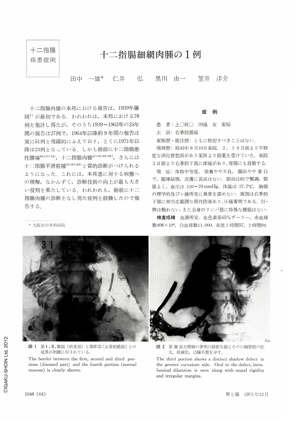
十二指腸肉腫の本邦における報告は,1939年藤岡1)が最初である.われわれは,本邦における78例を集計し得たが,そのうち1939~1963年の25年間の報告は27例で,1964年以降約9年間の報告は実に51例と飛躍的にふえており,とくに1971年以降は23例となっている.しかも術前に十二指腸悪性腫瘍68)72)74),十二指腸肉腫47)50)85)92),さらには十二指腸平滑筋腫81)87)89)と質的診断がつけられるようになった.これには,本疾患に対する病態への理解,なかんずく,診断技術の向上が最も大きい役割を果たしている.われわれも,術前に十二指腸肉腫の診断をなし得た症例を経験したので報告する.
A 39-year-old woman who visted our hospital with a complaint of pain in the right upper quadroant was preoperatively confirmed to harbor sarcoma of the duodenum as diagnosed by hypotonic duodenography. Not only the pancreas head which was already involved at the time of operation but also duodenum were excised. The neoplasm had intramurally spread into the anterior and posterior wall of all the portions of the duodenum, showing a mixed picture of hypertrophy, thinning, necrosis and perforation of the duodenal wall. Histologically it proved to be reticulum cell sarcoma. The patient died 6 months later on account of perforation of metastatic lesions of the intestine.
Discussion has been made on 78 such cases hitherto published in Japan with special reference to its literature. It was Fujioka who made the first report (1939). With the progress made in the field of hypotonic duodenography or selective angiography of the abdominal cavity the number of report is steadity increasing; within 2 years since 1971 as many as 23 cases. The age group 50 to 59 was most often affected, the male outnumbering the female with a ratio of 44 : 33. Signs and symptoms most frequently seen were anemia (76 per cent), palpation of the tumor (71 per cent), melena (40 per cent) and abdominal pain (37 per cent). Symptoms referrable to occulusion were few. The patterns of tumor development were extraluminal in 66 per cent, intraluminal in 20 and intramural in 14 per cent. The tumor often reached the size of a child's fist. The neoplasm developed in the first portin of the doudenum in 16 per cent; 52 per cent in the second and 13 per cent in he third portion, with 19 per cent elsewhere. Histologically, leiomyosarcoma formed a large majority (83 per cent) followed by reticulum cell sarcoma (10 per cent), angiosarcoma (3 per cent) and fibrosarcoma (1 per cent). X-ray findings naturally vary according to the patterns of tumor development, its size and sit of origin and its histologic types. Reticulum cell sarcoma mostly develops within the duodenal wall, and its x-ray findings are variegated, as in the present case, according to the gross findings such as loss of normal mucosa, shadow defect, deformity of the contour associated with intraluminal dilatation, mural rigidity, irregular outline, ulceration and perforation.

Copyright © 1973, Igaku-Shoin Ltd. All rights reserved.


