Japanese
English
- 有料閲覧
- Abstract 文献概要
- 1ページ目 Look Inside
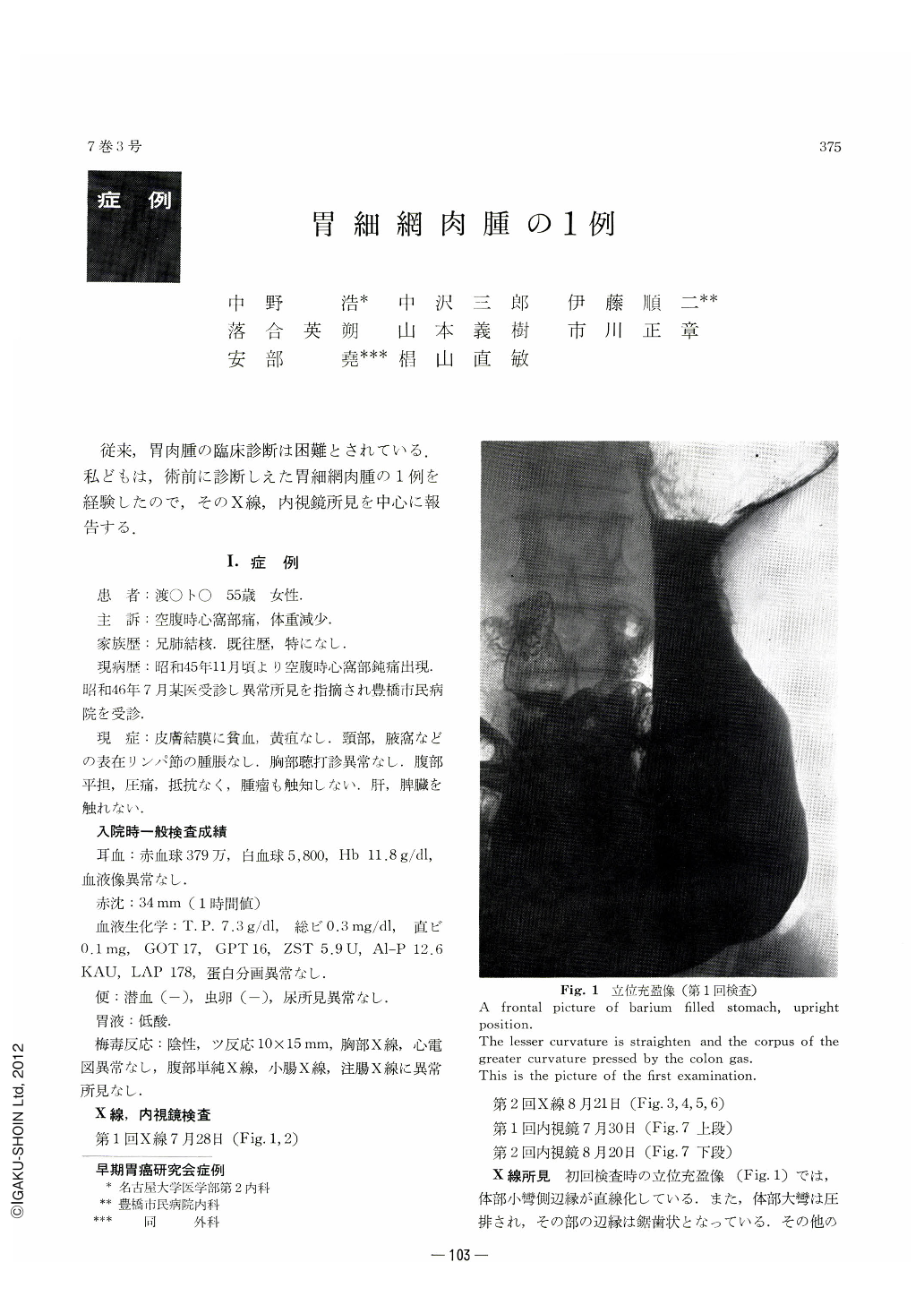
従来,胃肉腫の臨床診断は困難とされている.私どもは,術前に診断しえた胃細網肉腫の1例を経験したので,そのX線,内視鏡所見を中心に報告する.
A 55-year-old woman, with a history of a months long hunger pain in the epigastrium, had lost 7 kg in weight. No abnormality was found both in the status praesens and routine clinical examinations.
X-ray and endoscopic findings of the stomach are summarized as follows:
1. Multiple masses of peculiar shape were found on the posterior wall of the gastric body, converging to a central ulcer in a four-leaf-clover fashion. They were round and plateau-like with smooth borders. Almost all of the surface of the elevations were occupied by shallow, concentric depressions with their rims smooth and sharply circumscribed. These depressions with white coats on their surface were identified as erosions endoscopically. The borders of the masses excepting eroded parts were of normal but distended mucosa, presenting a characteristic finding of a submucosal tumor.
2. In a short time the forms of the lesions underwent a remarkable change; the masses increased in diameter and the number and form of each mass could be separately recognized.
3. Symmetrical ulcer formation was observed both in the center of the elevated masses on the posterior wall and just at the opposite side on the anterior wall.
4. Any definite proof suggesting of cancer was not encountered in all of the examinations.
These results led us to a clinical diagnosis of malignant lymphoma of the stomach.
The final diagnosis after surgical and pathological explorations was reticulum cell sarcoma with scars of mutiple ulcers. Histologically, the proliferation and infiltration of tumor cells were limited to the mucosa and submucosa, but those cells were also found in the regional lymph nodes.
According to the literature on reticulum cell sarcoma of the stomach, macroscopic and radiological findings of this case are considered to be quite typical of it.

Copyright © 1972, Igaku-Shoin Ltd. All rights reserved.


