Japanese
English
- 有料閲覧
- Abstract 文献概要
- 1ページ目 Look Inside
食道の良性腫瘍は比較的まれであり,自覚症状にも乏しく,上部消化管の検索時偶然に発見されるものも少なくない.Granular cell tumorは1926年のAbrikossoffの報告1)以来今日まで1,000例近くの報告があるが,消化管,なかでも食道に発生したものはわずか19例が報告されているにすぎない.
今回,われわれは内視鏡施行時の生検によって食道のgranular cell tumorと診断できた症例を経験したので多少の文献的考察を加えて報告する.
The patient, a 44 year-old man, had a complaint of dysphagia and was examined in our clinic.
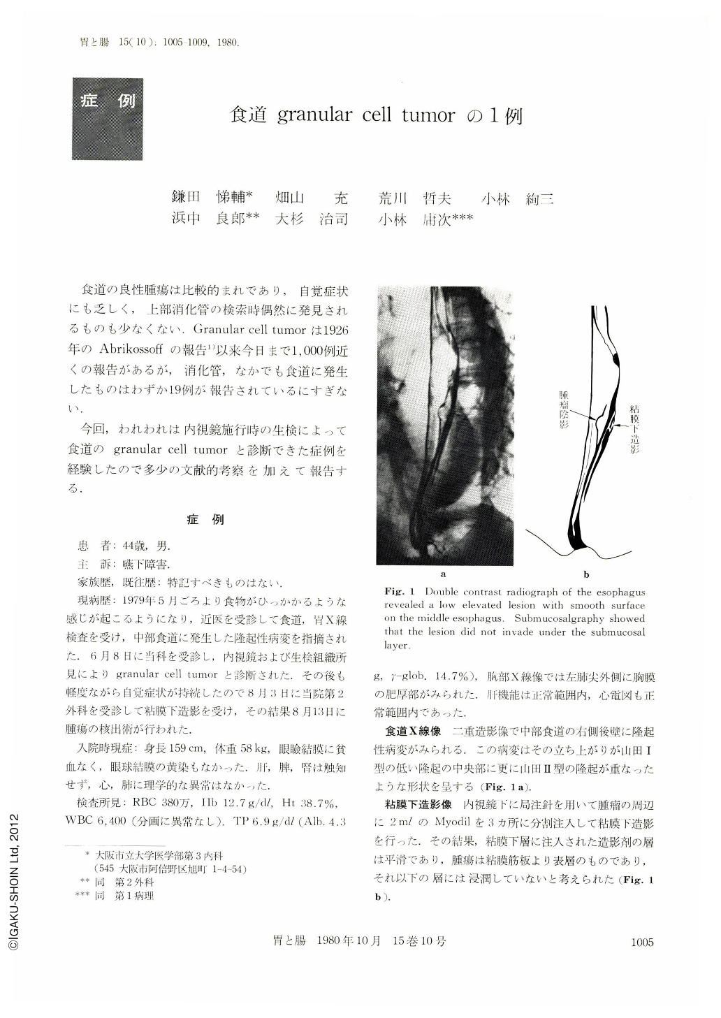
Roentgenologic and endoscopic findings demonstrated a smooth, yellowish-white and well-defined elevation at the left side of the middle esophagus. Biopsy specimens taken from the lesions disclosed the tumor cells with intracytoplasmic eosinophilic granules growing in the subepithelial layer and the overlying epithelium accompanied with pseudoepitheliomatous hyperplasia.
The ultrastructural findings of the present case were as follows.: 1) Tumor cells had numerous acidophilic cytoplasmic granules which were double membranebound and contained granular material, myelin substance and multivesicular bodies. 2) These tumor cell clumps were surrounded by basement membranes.
So, these morphological studies revealed granular cell tumor and suggested Schwann cell histogenesis.
The lesion were enucleated after being comfirmed to localize under the tunica propria mucosae on the roentgenogram with injected Myodil into the submucosal layer.
Resected tumor was yellowish-white, measuring 25×17×7 mm. And its lesion was comfirmed histologically to have no invasion into the submucosal layer.
To our knowledge only nineteen cases of this tumor were reported in the world literature and the present case was its 20 th.
Copyright © 1980, Igaku-Shoin Ltd. All rights reserved.