Japanese
English
- 有料閲覧
- Abstract 文献概要
- 1ページ目 Look Inside
囊腫の大きさが直径20cmに及ぶ先天性総胆管囊腫を経験したので,若干の考察を加えて報告する.
The patient: a 30-year-old man.
Chief complaint: Sensation of fullness in the abdomen.
Present illness: Since October 1980 the patient complained of sensation of fullness in the abdomen. As he had additionally slight pain in the abdomen, he visited Saeki Clinic. X-ray examination of the stomach was suggestive of apancreatic cyst, so that he was referred to our department for a complete checkup. All through the course of his illness no anemia or jaundice was noticed.
Present status: The patient was a man of moderate build and well-nourished. There was no icterus. A cystic tumor of smooth surface about 16 by 20 cm was palpated in the right upper part of the abdomen.
Laboratory examination of the blood showed that GOT was 265U, GPT 343U, total bilirubin 2.0mg/dl (DB 1.3mg/dl), LAP 445U, ALP 1, 724U, LDH 412U, serum amylase 274U, urine amylase 1983 U. CEA was 3.1mg/ml and a-fetoprotein was negative.
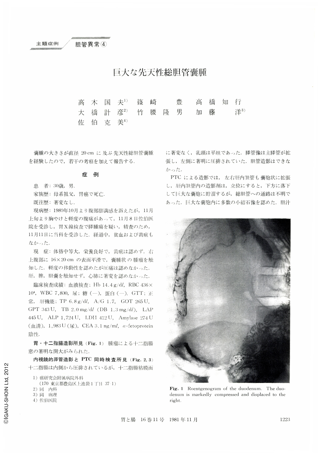
Opacification of the stomach and duodenum: The duodenal window was markedly enlarged.
ERCP: Though theduodenum itself was displaced and compressed, the mucosal surface was normal and the papilla was flat. The main pancreatic duct was dilated and displaced to the left in a high degree.
Cholangiography was unsuccessful.
PTC: Intrahepatic bile ducts were dilated as well like cysts, and the contrast medium within the intrahepatic ducts dropped down when the patient was ordered to stand upright, forming a mass within the gigantic cyst. Many small stones were recognizedwithin it.
Simultaneous examination with ERCP and PTC revealed a gigantic cyst and its relationship with intrahepatic ducts and pancreatic ducts. The cystmeasured 30 by 20cm.
On December 24, 1980, the cyst was removed along with cholecystectomy and cholangio-jejunal anastomosis. The resected cyst measured 20 by 20cm. About 2,500ml of bile was aspirated during the operation.
The wall of the cyst was fibrous and 0.5 to 1.0mm thick. There was no tumor within the inner surface. During the operation the part of confluence with the cyst and the main pancreatic duct remained unclarified.
Histologically, papillary projection was seen here and there on the inner surface of the cyst, but no diagnosis of cancer was made.
There are reports of congenital cyst of the common bile duct associated with cancer, and in our case the epithelia of the inner surface of the cystic wall showed slight atypicality. We believe such a cyst should be removed by all means in view of possible prospects of malignancy.
Copyright © 1981, Igaku-Shoin Ltd. All rights reserved.