Japanese
English
- 有料閲覧
- Abstract 文献概要
- 1ページ目 Look Inside
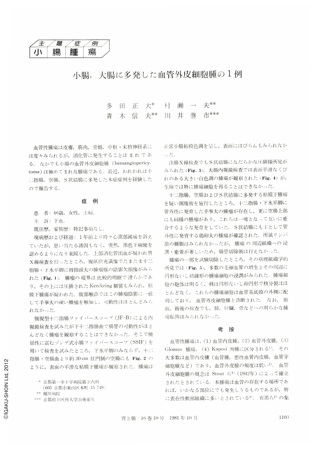
血管性腫瘍は皮膚,筋肉,骨髄,中枢・末梢神経系には度々みられるが,消化管に発生することはまれである.なかでも小腸の血管外皮細胞腫(hemangiopericytoma)は極めてまれな腫瘍である.最近,われわれは十二指腸,空腸,S状結腸に多発した本症症例を経験したので報告する.
A 46-year-old woman complaining of melena was admitted to our hospital. Barium meal study revealed a circumscribed tumor at the horizontal portion of the duodenum. Although enteroscopy with a conventional push-type duodenoscope failed because of the lack of the movement of the invaded duodenum, observation with a sonde-type small intestinal fiberscope (SSIF) which was designed to be much more slender and soft was successfully performed. Not only the lesion of the duodenum was revealed by x-ray study, but another lesion was clearly detected at the upper jejunum. They looked like submucosal tumors; the surface was smooth and there were no erosions.
Colonoscopy revealed another tumor which was semi-edunculated, white and hard, at the sigmoid colon. By operation, these tumors were histologically diagnosed as hemangiopericytoma arising from the duodenum, jejunum and sigmoid colon.
Hemangiopericytoma is a rare tumor and most have been found in the superficial soft tissues. Hemangiopericytoma in the jejunum is extremely rare and has not been repotted in Japan. This case is the first to be reported in Japan and its endoscopic pictures are the first to be shown in the world.

Copyright © 1981, Igaku-Shoin Ltd. All rights reserved.


