Japanese
English
- 有料閲覧
- Abstract 文献概要
- 1ページ目 Look Inside
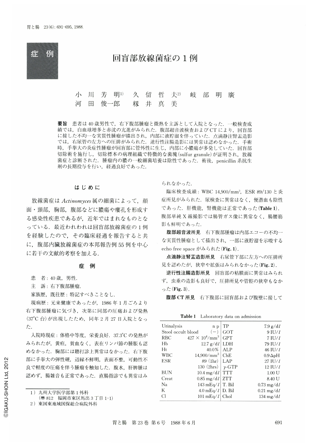
要旨 患者は40歳男性で,右下腹部腫瘤と微熱を主訴として入院となった.一般検査成績では,白血球増多と赤沈の亢進がみられた.腹部超音波検査およびCTにより,回盲部に接した不均一な実質性腫瘤が描出され,内部に液貯溜を伴っていた.点滴静注腎盂造影では,右尿管の左方への圧排がみられた.逆行性注腸造影には異常は認めなかった.手術時,手掌大の炎症性腫瘤が回盲部に管外性に生じ,内部に小膿瘍が多発していた.回盲部切除術を施行し,切除標本の病理組織で特徴的な菌塊(sulfur granule)が証明され,放線菌症と診断された.腫瘤内の膿の一般細菌培養は陰性であった.術後,penicillin系抗生剤の長期投与を行い,経過良好であった.
A 40 year-old man was admitted to our hospital with a low fever, and a tumor on the right side of the lower abdomen. Routine laboratory examination showed leucocytosis and increased ESR (Table 1). Ultrasonography demonstrated a solid mass of heterogeneous density with internal echo-free space suggesting fluid collection (Fig. 1). It seemed that the mass was located in the ileocecal region and was adhering to the abdominal wall on body CT (Fig. 4). Drip infusion pyelography revealed the right lower ureter had shifted to the left (Fig. 2). Barium enema disclosed no significant abnormality of the ileocecal mucosa (Fig. 3). On laparotomy, a fist sized inflammatory mass, showing extraluminal growth, was found adjacent to the ileocecal region, and it contained multiple small abscesses. The mass was removed by ileocecal resection (Fig. 5), and end-to-end anastomosis was performed. Histologically, sulfur granules in the resected specimen brought definite diagnosis of actinomycosis (Fig. 6). Bacteriological study of the pus in the abscesses was negative. After the operation, penicillin was administered for a long term, and the wound healed properly.
Copyright © 1988, Igaku-Shoin Ltd. All rights reserved.