Japanese
English
- 有料閲覧
- Abstract 文献概要
- 1ページ目 Look Inside
- サイト内被引用 Cited by
食道癌の検診は発病率が低いことや,X線検査での微細病変の描出が容易でないなどの理由からあまり実施されていない.
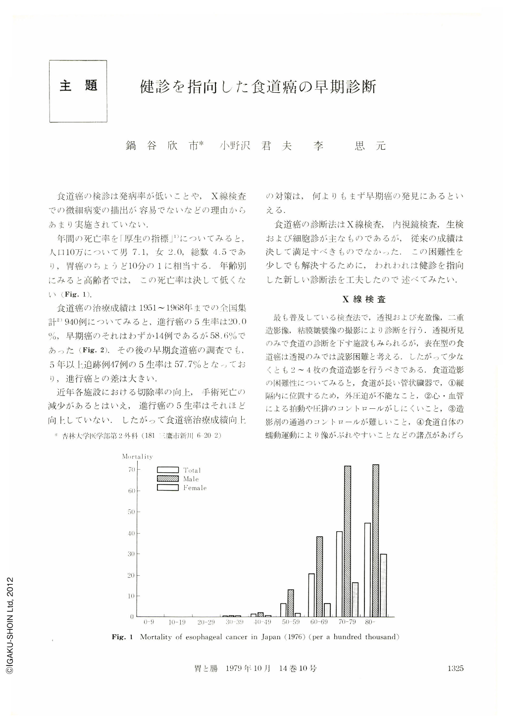
年間の死亡率を「厚生の指標」1)についてみると,入口10万について男7.1,女2.0,総数4.5であり,胃癌のちょうど10分の1に相当する.年齢別にみると高齢者では,この死亡率は決して低くない(Fig. 1).
Esophageal cancer has been mainly diagnosed by X-ray examination, esophagoscopy, biopsy and cytology. For the viewpoint of the early detection for the esophageal cancer, the results up to now are far from satisfactory.
In order to resolve the difficulty for early detection, we have developed a new method on the medical examination. That is as follow;
1, Serial esophagogram
One makes the patient swallow 40 ml barium at one time, and takes five pictures of it continuously at 1, 3, 5, 7 and 9 seconds. This method is of great use for us to diagnose fine lesions because we can get such pictures as filling, double contrast and relief in succession.
2, Esophageal brush cytology with capsule
The round shaped urethane-foam brush with thread is used as an instrument. The brush is enclosed in a capsule for administration. One makes the patient swallow it down to the stomach. The capsule melts 5 minutes later and the brush becomes to the original state. When pulled up the thread, some esophageal mucosal cells can be collected because of rubbing the esophagus by the brush. The method is simple and safe, and gives almost no pain to the patient. It is also useful for the screening in esophageal cancer of the outpatients and for the mass examination of esophageal cancer.
The results of examination patients with esophageal cancer are positive in 35 cases, doubtful in 12 and negative in 3. Among them, the positive rate of 5 superficial types was 100%.
We would like to emphasize that the use of the above mentioned method greatly contributes to the early detection of esophageal cancer.

Copyright © 1979, Igaku-Shoin Ltd. All rights reserved.


