Japanese
English
- 有料閲覧
- Abstract 文献概要
- 1ページ目 Look Inside
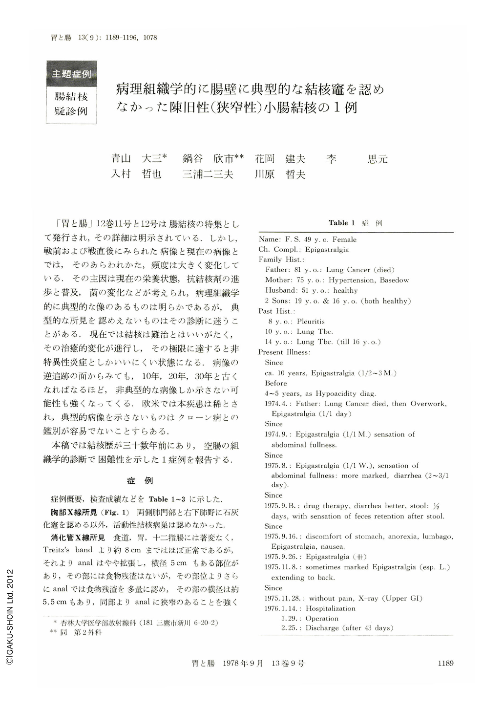
「胃と腸」12巻11号と12号は腸結核の特集として発行され,その詳細は明示されている.しかし,戦前および戦直後にみられた病像と現在の病像とでは,そのあらわれかた,頻度は大きく変化している.その主因は現在の栄養状態,抗結核剤の進歩と普及,菌の変化などが考えられ,病理組織学的に典型的な像のあるものは明らかであるが,典型的な所見を認めえないものはその診断に迷うことがある.現在では結核は難治とはいいがたく,その治癒的変化が進行し,その極限に達すると非特異性炎症としかいいにくい状態になる.病像の逆追跡の面からみても,10年,20年,30年と古くなればなるほど,非典型的な病像しか示さない可能性も強くなってくる.欧米では本疾患は稀とされ,典型的病像を示さないものはクローン病との鑑別が容易でないことすらある.
本稿では結核歴が三十数年前にあり,空腸の組織学的診断で困難性を示した1症例を報告する.
A 49-year-old woman was admitted with complaints of left epigastralgia, anorexia and nausea. At the ages of 8, 10 and 14 until 16, she had suffered from pleurisy and lung tuberculosis. Since one year and seven months after the death of her father she had complained of epigastralgia.
Routine barium meal X-ray studies revealed jejunal stenoses in two places. At each site small ulcers 2~3 mm in diameter were suspected. At the oral side of the upper jejunum were depicted dilatation 5.5 cm long and many food particles.
Jejunal tuberculosis was indicated by the gross findings of the resected specimens of the jejunum as well as by the radiological findings. Although no caseating granuloma was found in any of the walls of the resected jejunal segments, the lymph node of the mesenterium showed typical caseating granuloma.
Diagnosis of lesions in the small and large intestine must be very carefully made.

Copyright © 1978, Igaku-Shoin Ltd. All rights reserved.


