Japanese
English
- 有料閲覧
- Abstract 文献概要
- 1ページ目 Look Inside
特異な臨床像と病理学的特徴をもつ消化管のVillous adenomaは本邦では比較的稀なものとされ,その好発部位は直腸と言われている.最近,われわれは胃幽門前庭部に,大腸にみられるVillous adenomaに類似したⅡa集簇型胃癌とBorrmann2型胃癌が衝突した1例を経験したので報告する.
Our current report is based on a case of collision carcinoma of the stomach occurred on the antrum, in which Borrmann 2 Type coexisted with conglomerated Ⅱa Type lesion which showed similarity to villous adenoma of the large intestine in its developing course.
The patient: 65-year-old female. Chief complaints: dull pain in the upper abdomen since two months before. Physical examination and laboratory data: no particular abnormalities.
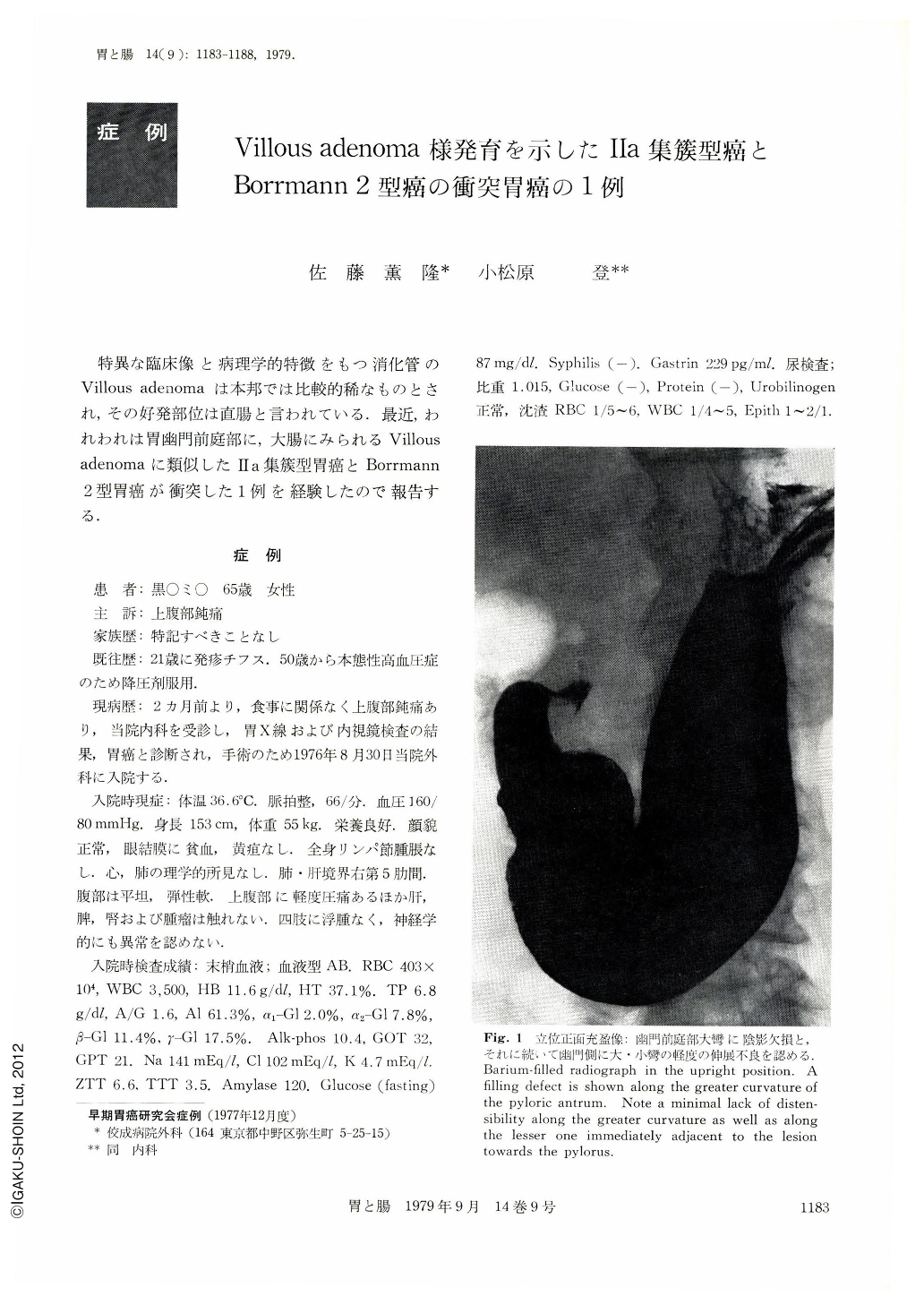
Barium and endoscopical examination of the stomach revealed an elevated lesion along the greater curvature of the antrum characterized by low prominence, oval configuration with a structure of 4.5×3.5 cm in diameter: immediately adjacent to that lesion, towards its prepyloric side was another conglomeration of elevated lesions of varied size, apparently softer and lower in height. Histological study of the biopsy specimen confirmed the character of papillo-tubular adenocarcinoma besides an accurate diagnosis of Borrmann 2 type for the former and conglomerated Ⅱa Type for the latter. The gastrectomy was performed on Sept. 8, 1976 according to the Billroth I method and the patient is still alive after having transcurred 2 years and 3 months since the operation. The resected specimen of the stomach shows two lesions: one Borrmann 2 Type of 4.0×3.0cm in diameter, localized 5cm towards the oral side of the pyloric ring along the greater curvature; the other lesion of conglomerated nodules of varied size, localized adjacently of the former, encircling the pre-pylorus (9.0×2.2cm)
Histologically Borrmann 2 Type lesion resulted to be papillo-tubular adenocarcinoma (depth of invasion p.m. INF, β, (++), v (-) 1/14, stage Ⅱ) and the conglomerated nodular lesion turned out to be atypical glands proliferated in villous pattern with broad base; its grade of atypicity was low in most parts of the lesion with resemblance to atypical epithelial cells, but partly the histology revealed the carcinoma of it.
This lesion has similarity with villous adenoma of the large intestine, nevertheless it should be considered as a conglomerated Ⅱa Type gastric carcinoma (depth of invasion m.), highly clifferentiated. Also the current case should be categorized as one of collison carcinoma of the stomach since the two lesions are clearly delimited the one from the other.

Copyright © 1979, Igaku-Shoin Ltd. All rights reserved.


