Japanese
English
- 有料閲覧
- Abstract 文献概要
- 1ページ目 Look Inside
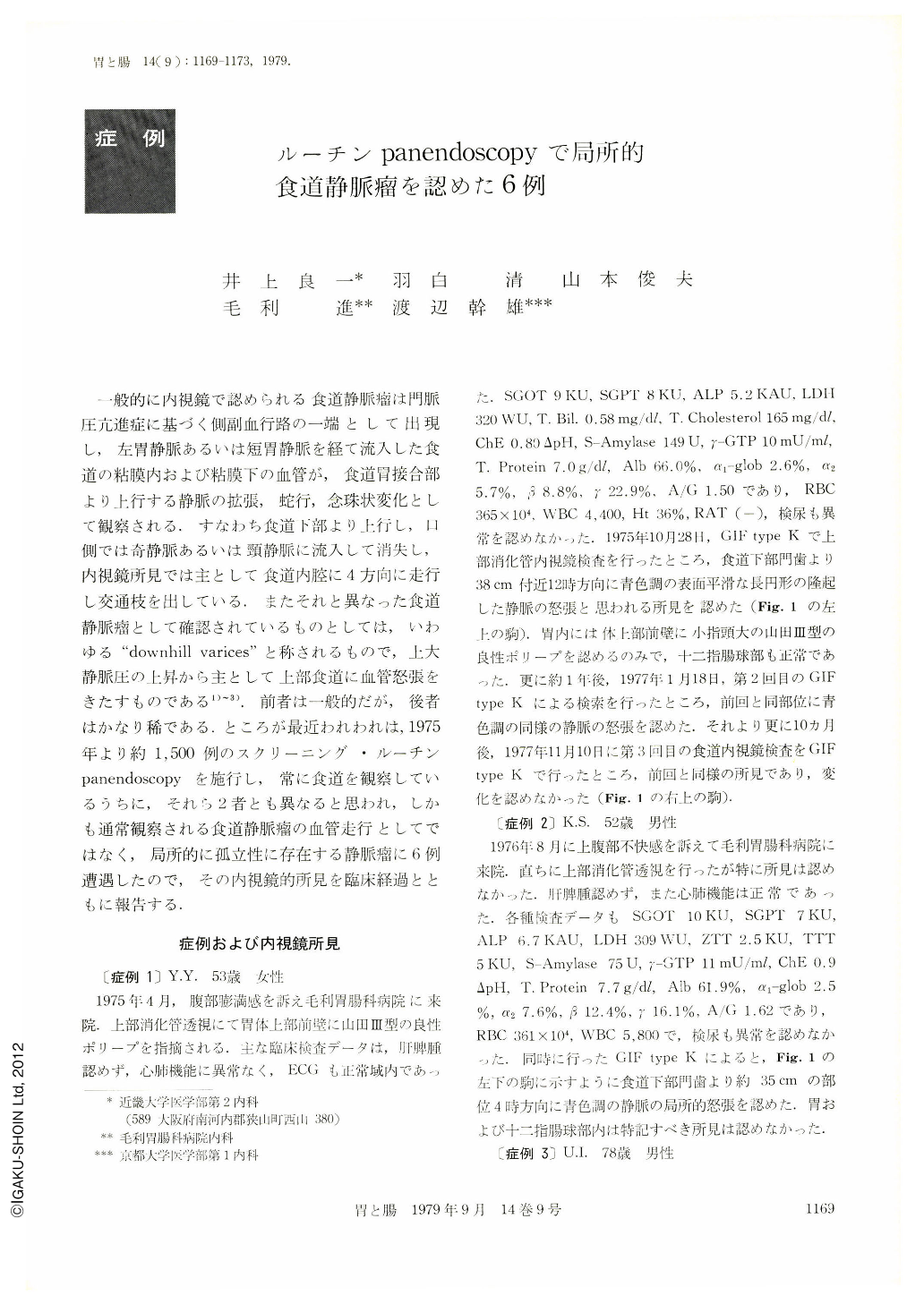
一般的に内視鏡で認められる食道静脈瘤は門脈圧亢進症に基づく側副血行路の一端として出現し,左胃静脈あるいは短胃静脈を経て流入した食道の粘膜内および粘膜下の血管が,食道胃接合部より上行する静脈の拡張,蛇行,念珠状変化として観察される.すなわち食道下部より上行し,口側では奇静脈あるいは頸静脈に流入して消失し,内視鏡所見では主として食道内腔に4方向に走行し交通枝を出している.またそれと異なった食道静脈瘤として確認されているものとしては,いわゆる“downhill varices”と称されるもので,上大静脈圧の上昇から主として上部食道に血管怒張をきたすものである1)3).前者は一般的だが,後者はかなり稀である.ところが最近われわれは,1975年より約1,500例のスクリーニング・ルーチンpanendoscopyを施行し,常に食道を観察しているうちに,それら2者とも異なると思われ,しかも通常観察される食道静脈瘤の血管走行としてではなく,局所的に孤立性に存在する静脈瘤に6例遭遇したので,その内視鏡的所見を臨床経過とともに報告する.
Varices of the esophagus are a frequent occurence in the portal hypertention caused by liver cirrhosis. Other varices of the upper esophagus are very unusually caused by obstruction of surperior vena cava or thyroid tumor. These varices are called “downhill varices”. The author performed over 1500 routine panendoscopic examinations and found the localized varices of the esophagus in 6 patients. These localized varices are distinctly different from both esophageal varices. Because these 6 cases have neither liver diseases nor congestive heart diseases. They also have no thyroid diseases. The endoscopic ficdings of the localized varices are local or solitaly venous dilatation, which are not continued to other esophageal mucosal veins. Their color is almost blue tone. Their surface is almost smooth. The etiology of the localized varices is not known, because they may not be associated definite with diseases.

Copyright © 1979, Igaku-Shoin Ltd. All rights reserved.


