Japanese
English
- 有料閲覧
- Abstract 文献概要
- 1ページ目 Look Inside
症 例
患 者:63歳の男子
主 訴:上腹部不快感
家族歴:特記することはない
既往歴:58歳時,虫垂切除術施行
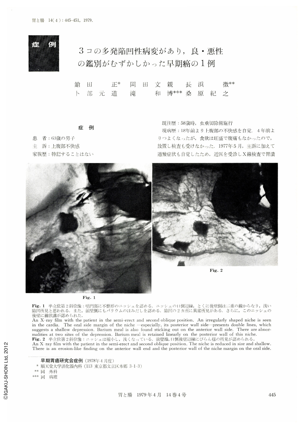
現病歴:18年前より上腹部の不快感を自覚.4年前よりつよくなったが,食欲は旺盛で腹痛もなかったので,放置し検査も受けなかった.1977年5月,主訴に加えて過酸症状も自覚したため,近医を受診しX線検査で胃潰瘍と診断された.薬物療法で症状はすぐよくなった.しかし,9月に症状が再発したので当科で精査した.3カ所に潰瘍性病変があり,おのおの早期癌,早期癌の疑い,および良性潰瘍と診断した.
A 63-year-old man with a complaint of discomfort in the upper abdominal region for 18 years was suspected of having type Ⅱc and Ⅲ early gastric cancers, with an ulcer scar as the result of detailed preoperative examinations Three depressed lesions were thus diagnosed.
Detailed histologic examinations of the totally resected stomach disclosed that there was a 1.8×1.3 cm lesion of Ⅱc+Ⅱa early cancer (tubular adenocarcinoma, with an invasion of sm) in the antrum, and that in the cardia a type Ⅲ early cancer (tubular adenocarcinoma, with an invasion of sm in a part of serial sections) occurred focally (1.9×0.5 cm) surrounded by the regenerated epithelium in a part of the margin of an active ulcer (Ul-Ⅳ) located in the center of a linear ulcer 4.7 cm in length.
Because the case mostly satisfied the Nakamura's criteria for the diagnosis of ulcer-cancer, canceration of ulcer was considered. This lesion had preoperatively been caught hold of by roentgenography and also by endoscopy, but cancer in this part could not have been demonstrated by directed biopsy. This impressed us with the difficulty in biopsy diagnosis of type Ⅲ early cancer. These lesions were quite independent of each other, presenting nothing suggestive of intramural metastasis and invasion.
The shallow depressed lesion, 1.5×1.2 cm, located in the angular incisure, was a scar of UI-Ⅲ, with intestinal epithelial cells coexistent with Paneth cells in the deep mucosal layer; and the glandular epithelium constituting the upper half of the mucosa was a lesion made up of intestinal epithelial atypical cells and the regenerated epithelium continuous from the adjacent area, which could not be diagnosed as cancer yet. It was such a borderline lesion that even the pathologists were divided in opinion. This atypical epithelium could not have been diagnosed by the detailed preoperative examinations but as a scar of benign ulcer.
It remains problematic whether this lesion shows a profile of canceration of ulcer or is a mere complication of atypical epithelium by ulcer. Today, when canceration of atypical epithelium cannot entirely be ruled out, the diagnosis of whether a depressed lesion is benign or malignant requires data from more cases, and thus it remains a theme for future studies.

Copyright © 1979, Igaku-Shoin Ltd. All rights reserved.


