Japanese
English
- 有料閲覧
- Abstract 文献概要
- 1ページ目 Look Inside
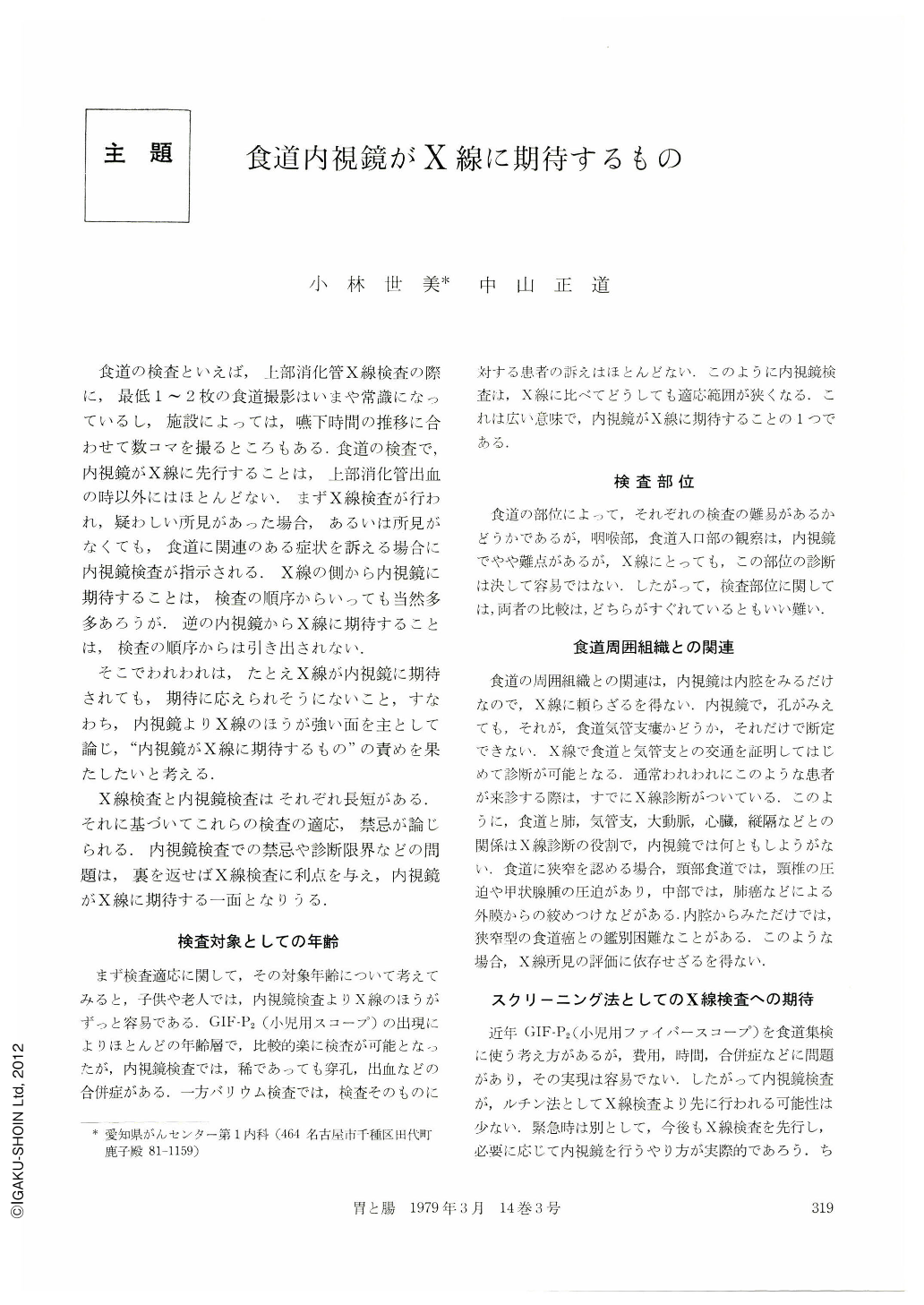
食道の検査といえば,上部消化管X線検査の際に,最低1~2枚の食道撮影はいまや常識になっているし,施設によっては,嚥下時間の推移に合わせて数コマを撮るところもある.食道の検査で,内視鏡がX線に先行することは,上部消化管出血の時以外にはほとんどない.まずX線検査が行われ,疑わしい所見があった場合,あるいは所見がなくても,食道に関連のある症状を訴える場合に内視鏡検査が指示される.X線の側から内視鏡に期待することは,検査の順序からいっても当然多多あろうが.逆の内視鏡からX線に期待することは,検査の順序からは引き出されない.
そこでわれわれは,たとえX線が内視鏡に期待されても,期待に応えられそうにないこと,すなわち,内視鏡よりX線のほうが強い面を主として論じ,“内視鏡がX線に期待するもの”の責めを果たしたいと考える.
It is unusual that endoscopy does expect X-ray examination in the diagnosis of esophageal disease, because endoscopic examination is usually performed following X-ray examination. Therefore, we herein will discuss mainly on the advantages of X-ray examination compared to endoscopy in certain respects.
First of all, X-ray examination is more easily and safely carried out in pediatric and elderly patients than endoscopy, because complications may be more often encountered with endoscopy in those patients.
Secondly, X-ray examination is more beneficial to obtain informations on the neighboring organs or tissues of the esophagus.
Thirdly, X-ray examination plays an important role as a screening method which is expected to reveal even a very small abnormality.
Fourthly, X-ray examination reveals configuration of the entire esophagus even in patients with advanced carcinoma of the esophagus through which an endoscope can not pass down distally. In our series of esophageal carcinoma, the endoscope could pass through the lesion in only 25%. However, the development of GIF P2 (pediatric gastrointestinal fiberscope) could improve up to nearly 70% in passing through a stenotic lesion. X-ray examination is also more beneficial in detecting achalasia, scleroderma, webs in the upper esophagus and mild hiatal hernia.
Fifthly, X-ray examination is only reliable diagnostic method at the institutions without endoscopic facilities.

Copyright © 1979, Igaku-Shoin Ltd. All rights reserved.


