Japanese
English
- 有料閲覧
PPV購入案内
1,320円 (1,200円+税10%) こちらは、1063ページから1066ページの文献です。購入には医書.jp本会員登録が必要です。
会員登録完了後に改めて上記「購入サイトへ」を選択してください。
または「購入サイトへ」選択後に「購入ログイン」、「新規会員登録」の順に進んでください。
会員登録について詳しくはをご確認ください。
- Abstract 文献概要
- 1ページ目 Look Inside
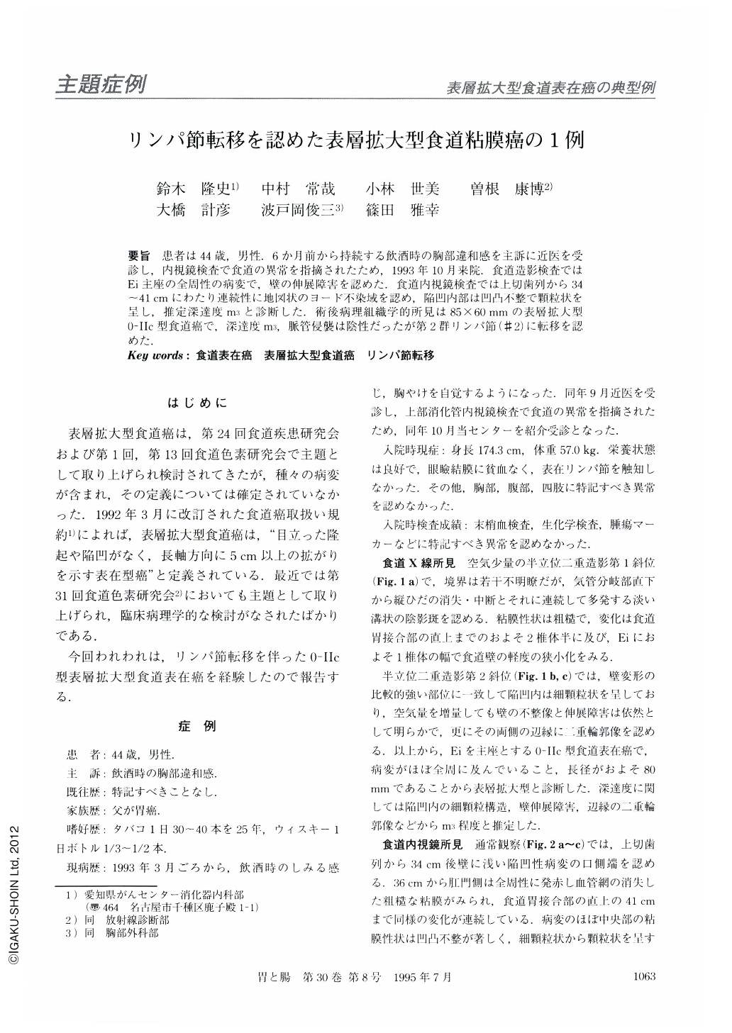
要旨 患者は44歳,男性.6か月前から持続する飲酒時の胸部違和感を主訴に近医を受診し,内視鏡検査で食道の異常を指摘されたため,1993年10月来院.食道造影検査ではEi主座の全周性の病変で,壁の伸展障害を認めた.食道内視鏡検査では上切歯列から34~41cmにわたり連続性に地図状のヨード不染域を認め,陥凹内部は凹凸不整で顆粒状を呈し,推定深達度m3と診断した.術後病理組織学的所見は85×60mmの表層拡大型0-Ⅱc型食道癌で,深達度m3,脈管侵襲は陰性だったが第2群リンパ節(#2)に転移を認めた.
A 44-year-old man was admitted to our hospital, with the complaint of anterior chest discomfort while drinking alcohol. Double contrast radiography of the esophagus revealed that mucosal folds disappeared at the proximal margin of abnormal barium coating and an annular lesion extended approximately 80 mm in length. Furthermore, the esophageal lumen showed a narrowing at the portion with the most irregular mucosal pattern despite sufficient air insufflation. Endoscopically, the lesion was seen as reddish, well-defined, and rough mucosa, which was not stained with iodine. The most granular and uneven changes of the esophageal mucosa were recognized at the center of the lesion in the lower esophagus. The diagnosis of superficial spreading carcinoma invading the muscularis mucosae (m3) was made. In addition, endoscopic ultrasonography revealed an enlarged intra-abdominal lymph node, suggesting a metastasis. Histological examination showed a well differentiated squamous cell carcinoma, 85 × 60 in size, with invasion of ms and lymph node metastasis (#2).
Copyright © 1995, Igaku-Shoin Ltd. All rights reserved.