Japanese
English
- 有料閲覧
- Abstract 文献概要
- 1ページ目 Look Inside
- サイト内被引用 Cited by
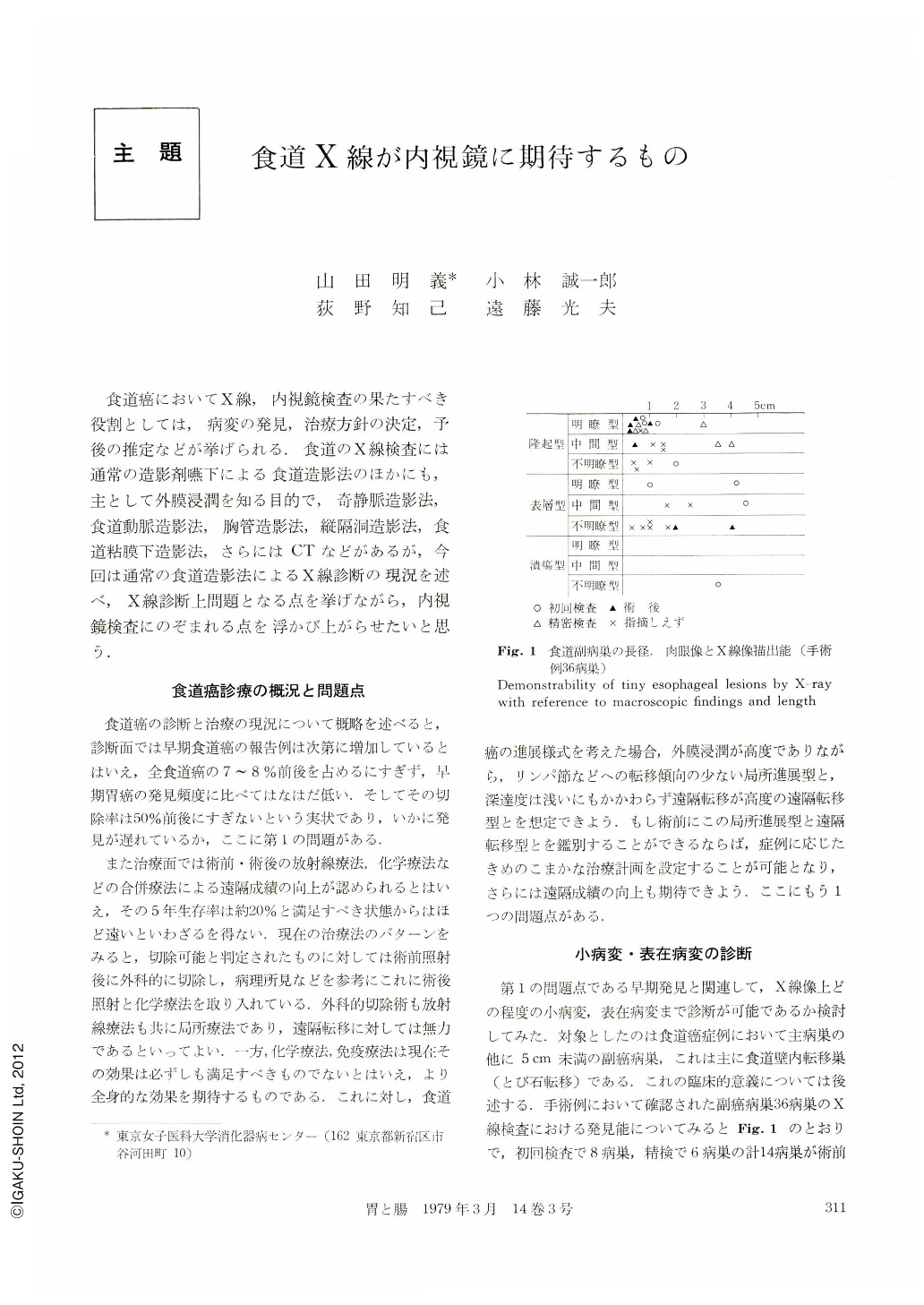
食道癌においてX線,内視鏡検査の果たすべき役割としては,病変の発見,治療方針の決定,予後の推定などが挙げられる.食道のX線検査には通常の造影剤嚥下による食道造影法のほかにも,主として外膜浸潤を知る目的で,奇静脈造影法,食道動脈造影法,胸管造影法,縦隔洞造影法,食道粘膜下造影法,さらにはCTなどがあるが,今回は通常の食道造影法によるX線診断の現況を述べ,X線診断上問題となる点を挙げながら,内視鏡検査にのぞまれる点を浮かび上がらせたいと思う.
Firstly, it is very important to find a tiniest possible lesion in esophageal carcinoma cases. We can find small lesions even 5 mm in diameter, if they are well-defined. However, if a lesion is ill-defined and of superficial or ulcerated type, a lesion even 20 mm in diameter might be overlooked.
In regard to depth of invasion of lesions which cannot be delineated by X-ray, it is usually limited within the muscularis mucosae. If the lesions invade the submucosal layer, we can delineate almost all of them except too tiny ones. The common macroscopical findings are changes of color tone, erosion-like changes and therefore endoscopy is superior to X-ray examination to getting these findings, for the latter is useful for finding only uneven lesions. The same may be said of the changes concomitant with esophageal varices and/or achalasia.
Secondly, it is also very important to establish diagnosis from which we can know indication for treatment and prognosis. As to local findings such as depth of invasion or invasion to the adventitia, X-ray examination should be applied, for it is convenient to catch a whole picture of the lesion.
On lymph node metastasis that influences prognosis, we can evaluate it only from local findings. We have discussed lymph node metastasis mainly in superficial carcinoma cases whose infiltration is limited within the submucosal layer. From the findings of the surface in superficially elevated type, the uneveness of the bottom of superficial depressed type, granular surface of superficially spread part of mixed type, skip metastatic lesions etc., we can evaluate the possibility of lymph node metastasis.
Finally, to detect and observe these tiny lesions, X-ray and endoscopy should always be utilized together.
Copyright © 1979, Igaku-Shoin Ltd. All rights reserved.