Japanese
English
- 有料閲覧
- Abstract 文献概要
- 1ページ目 Look Inside
胃体部腺領域に12コのcarcinoid病巣を有し,しかもargyrophil cell hyperplasiaを含む種々の発育段階を示す病巣を光顕的ならびに電顕的にも観察しえたので報告する.
症 例
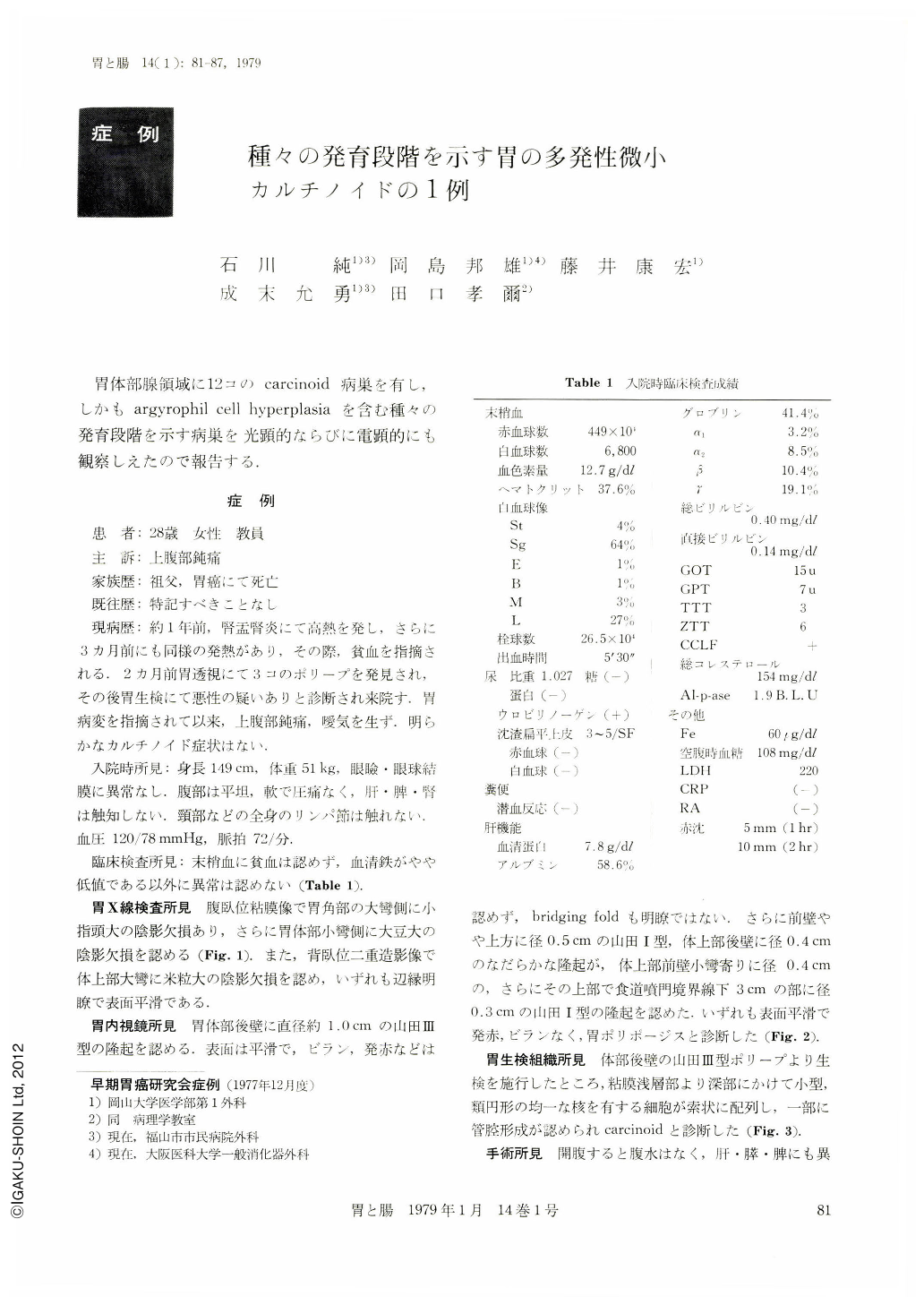
患 者:28歳 女性 教員
主 訴:上腹部鈍痛
家族歴:祖父,胃癌にて死亡
既往歴:特記すべきことなし
A case of multiple carcinoid tumor, initially the hyperplasia of gastric argyrophil cells, showing various developmental stages is presented. A 28-year-old woman complained of dull pain in the epigastrium without carcinoid symptoms. Subtotal gastrectomy was performed under the diagnoses of polyposis by fluoroscopic examination and of carcinoid tumor by the biopsy of the gastric mucosa. Macroscopically, one protruded lesion-Yamada's Type III, 1.0 cm in maximal diameter, and four Yamada's Type I lesions -0.6~0.4 cm in diameters were found, but scrupulous histological examination on the resected stomach revealed twelve carcinoid lesions in the mucosa of fundic gland region.
There were three mucosal and nine submucosal lesions showing positive argyrophil reaction but negative argentaffin reaction.
Adjacent to the carcinoid lesions, many tubules composed of one layered proper gastric glandular cells lying upon peripheral argyrophil cells were proliferated.
This feature was interpreted as argyrophil cell hyperplasia. Also, there were several minimal sized lesions, less than 0.1 mm in diameter immediately above the lamina muscularis mucosae. With the electron microscope, mitochondria and numerous round or oval granules, 200~400 mμ in diameters, entrapped by limiting membrane, were found in the cytoplasm of the tumor cells.
Furthermore, in the region of argyrophil cell hyperplasia, glandular cells having microvilli were connected with each other by desmosomes and formed obvious lumina. The cell having endocrine granules were situated at the outermost layer of the lumen and its outer surface was covered with the basement membrane.
These cells closely resembled ECL cell found in the gastrointestinal mucosa.
Regarding the histogenesis of the carcinoid tumor, the minimal sized lesion and the argyrophil cell hyperplasia seemed to be responsible for development of the tumor.
The patient has been well 10 months postoperatively and shows no sign of elevation of the serum serotonin level.
Copyright © 1979, Igaku-Shoin Ltd. All rights reserved.