Japanese
English
- 有料閲覧
- Abstract 文献概要
- 1ページ目 Look Inside
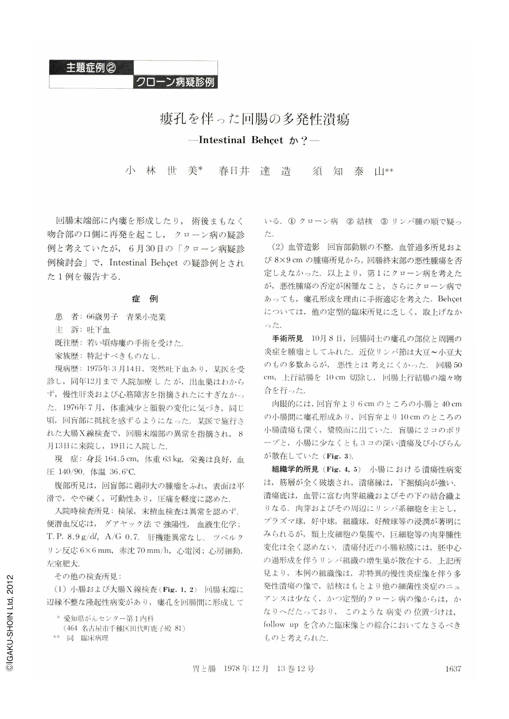
回腸末端部に内瘻を形成したり,術後まもなく吻合部の口側に再発を起こし,クローン病の疑診例と考えていたが,6月30日の「クローン病疑診例検討会」で,Intestinal Behçetの疑診例とされた1例を報告する.
症 例
患 者:66歳男子 青果小売業
主 訴:吐下血
既往歴:若い頃痔瘻の手術を受けた.
家族歴:特記すべきものなし.
現病歴:1975年3月14日,突然吐下血あり,某医を受診し,同年12月まで入院加療したが,出血巣はわからず,慢性肝炎および心筋障害を指摘されたにすぎなかった.1976年7月,体重減少と顔貌の変化に気づき,同じ頃,回盲部に抵抗を感ずるようになった.某医で施行された大腸X線検査で,回腸末端部の異常を指摘され,8月13日に来院し,19日に入院した.
A 66-year-old man was well until March 1975, when he developed hematemesis and melena. In July 1976, he developed right lower quadrant pain associated with a gradual weight loss.
On physical examination, he was noted to have a firm, mobile, egg-sized mass with a smooth surface and slight tenderness in the ileocecal region.
The patient was admitted to hospital for investigation.
Barium enema and barium meal follow-through studies revealed an irregular mucosal pattern in the terminal ileum with a fistula between the ileal loops. The radiological impression was probably Crohn's disease of the ileum.
However, because the possibility of tuberculosis or lymphoma could not be entirely ruled out. Ileocecal resection was performed. The resected specimen demonstrated multiple irregular ulcers and erosions with an internal fistula between the loops of the terminal ileum.
Histologically, these ulcers were of non-specific inflammatory process, with deeply undermined edges, penetrating through the muscularis. The floor of ulcer shows granulation tissues rich in vascularity, containing maked cellular infiltrates of lymphocytes, plasma cells, histiocytes, neurophils and eosinophils. Foci of lymphoid tissues with hyperplastic germinal centers were seen scattered through the ileum near the ulcers. The histological pictures were not typical of Crohn's disease. Tuberculosis and other infectious etiology were unlikely.
Postoperatively, ESR did not improve five weeks after operation. Four months later, he developed dearrhea and abdominal pain along the operative suture line. Barium enema at that time revealed a recurrent disease on the ileal side of the anastomosis. Since then, he has been treated with salazopyrin and predonisolone for one year and eight months. He is now in clinical remission
During the follow-up period, he was suspected of having Behçet's disease because of grossly undermined appearance of the ulcers. However, the patient has not yet shown other signs of Behçet's disease than occasional aphthous stomatitis which can also be a complication of Crohn's disease.
It has been reported that a trare occasions do typical of Behçet disease appear later than intestinal ulceration. A long-term follow-up should clarify the true nature of this patient's disease.

Copyright © 1978, Igaku-Shoin Ltd. All rights reserved.


