Japanese
English
- 有料閲覧
- Abstract 文献概要
- 1ページ目 Look Inside
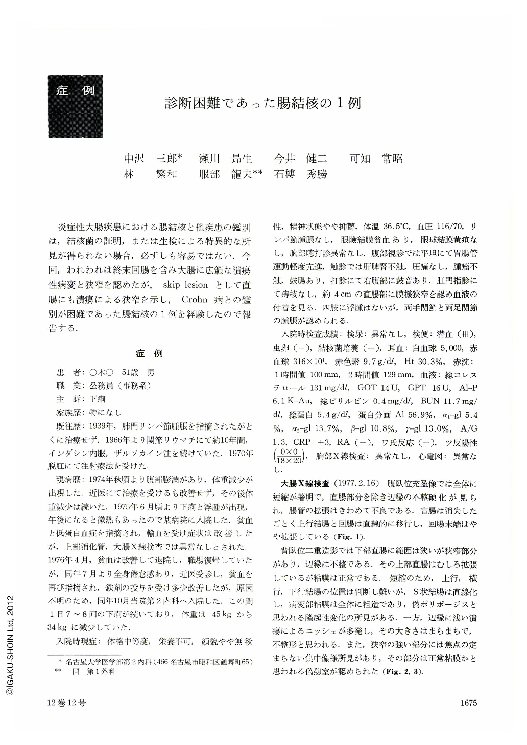
炎症性大腸疾患における腸結核と他疾患の鑑別は,結核菌の証明,または生検による特異的な所見が得られない場合,必ずしも容易ではない.今回,われわれは終末回腸を含み大腸に広範な潰瘍性病変と狭窄を認めたが,skip lesionとして直腸にも潰瘍による狭窄を示し,Crohn病との鑑別が困難であった腸結核の1例を経験したので報告する.
A 51 years old man was admitted to our hospital, complaining of diarrhea for two years. His past history showed swelling of hilar lymph node 37 years ago, rheumatic arthritis from 10 years ago and anal prolapsus six years ago.
He had anemia and hypoproteinemia. Stool examinations for occult blood were strongly positive. Cultures of stool and biopsy specimen were negative for tubercle bacillus.
Roentgenographic examination: Barium filled picture revealed marked shortening and stricture of the bowel and associated rectal stenosis. Double contrast picture showed ulcerative change with rough mucosal surface and mucosal convergence.
Endoscopic examination: An irregular-shaped ulcer and pseudopolyposis were seen of the descending colon. Stenosis of the rectum with bleeding was observed at a point 4 cm from anus. Biopsy showed only non-specific inflammation.
Pathological fingings: Subtotal colectomy was performed. Grossly resected specimen showed strong stricture whose wall was hypertrophic. Multiple ulcers and pseudopolyposis resembled cobblestone appearance. Rectal mucosa was normal, but there were ulcerations near the anal edge. Microscopically shallow ulcers spread broadly. Intestinal wall of the lesion was thickened by inflammatory granuloma and infiltrated by numerous lymphocytes and plasma cells.
Some epitheloid cell granuloma were observed, and on the other side tuberculous glanuloma with a tendency to caseation necrosis was observed. Characteristic tuberculous granuloma was observed in the lymph node.
This case was difficult to be differentiated from Crohn's disease, because it showed stricture, cobblestone-like finding and epitheloid cell granuloma. But the diagnosis was determined by detailed microscopic examination.

Copyright © 1977, Igaku-Shoin Ltd. All rights reserved.


