Japanese
English
- 有料閲覧
- Abstract 文献概要
- 1ページ目 Look Inside
腸管のう腫様気腫は腸管壁内に多数の小気胞が集簇して発生し,腸管があたかもブドウの房様を呈する比較的稀な疾患である.ときに気腫は腸間膜,大網,横隔膜などにも認められる.
本邦では1968年,江里ら1)が本疾患の148例を集計報告して以来,その後の5年間の著者らが文献上調査し得た37例を加えた計185例にすぎない.従来の報告1)2)では本疾患の発生部位は圧倒的に小腸に多いとされているが,われわれの調査した37例の少なくも12例(32%)では病変は結腸に認められており,結腸発生例の増加傾向がうかがわれた.しかし本疾患の内視鏡像についての報告はいまだ少ない.
A 52-year-old female was admitted to our hospital because of constipation, hematorrhea and hypogastric discomfort. Physical examinations disclosed no abnormal findings. The results of laboratory examinations were also normal.
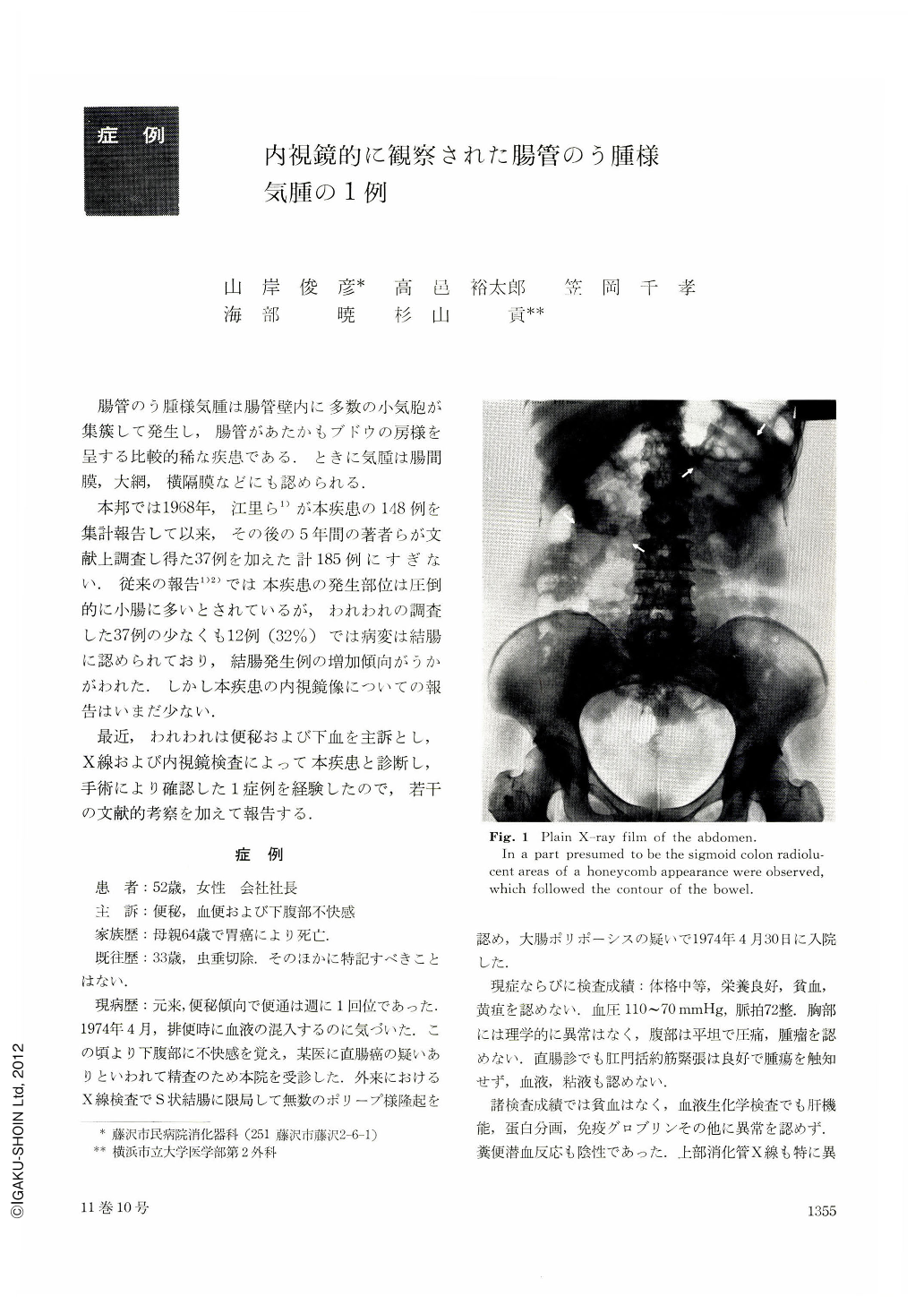
The plain X-ray film of the abdomen revealed a large amount of gas in the large intestine. In a part presumed to be the sigmoid colon radiolucent areas of a honeycomb appearance were observed, which followed the contour of the bowel. X-ray examination of the large intestine by double contrast method showed numerous polypoid lesions of various size with smooth surface in the middle part of the sigmoid colon. Barium filled picture revealed a filling defect in the shape of a holly leaf. Endoscopy disclosed numerous semispherical elevations size of various with smooth surface in the sigmoid colon. The color of such elevations was almost similar to the surrounding mucosa, but remarkable reddening was also present at places on the surface of some elevations. Furthermore, on the surface of the elevations submucosal vessels were observed. Partly the surface of the elevation was thin-walled and seemed to be transparent. From the above-mentioned results the case was diagnosed as pneumatosis cystoides intestinalis of the sigmoid colon, and operation was done. The sigmoid colon was fully movable under the state of so-called mobile sigma elongatum, and numerous cysts were observed subserosally. About 30 cm of the lesion on the sigmoid colon was resected and end-to-end anastomosis was performed. A number of cysts were noted subserosally and submucosally. Those lying close together looked like heaps of soap bubbles. The cyst was of spongy consistency and when pressed it burst with a pop discharging odorless gas. Histologically the burst inner wall of cyst consisted of a sigle layer of pavement cells and partly degeneration and decollement were observed. From the above the diagnosis was confirmed as pneumatosis cystoides intestinalis. Association with other gastrointestinal lesions was not observed.
The postoperative course was uneventful. She is well now with constipation being improved.
So far pneumatosis cystoides intestinalis found in Japan used to be in jejunum or the ileum, but investigations for the past five years indicated an increasing trend of occurrence in the colon.

Copyright © 1976, Igaku-Shoin Ltd. All rights reserved.


