Japanese
English
- 有料閲覧
- Abstract 文献概要
- 1ページ目 Look Inside
- サイト内被引用 Cited by
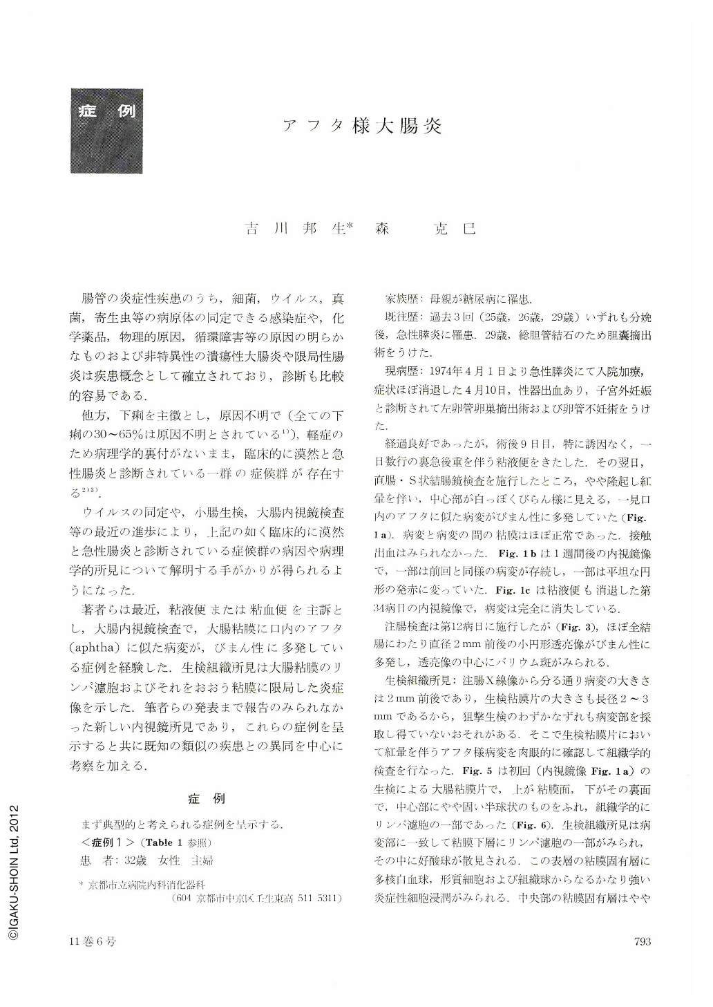
腸管の炎症性疾患のうち,細菌,ウイルス,真菌,寄生虫等の病原体の同定できる感染症や,化学薬品,物理的原因,循環障害等の原因の明らかなものおよび非特異性の潰瘍性大腸炎や限局性腸炎は疾患概念として確立されており,診断も比較的容易である.
他方,下痢を主徴とし,原因不明で(全ての下痢の30~65%は原因不明とされている1)),軽症のため病理学的裏付がないまま,臨床的に漠然と急性腸炎と診断されている一群の症候群が存在する2)3).
Numerous slightly elevated, pale lesions with red halos, resembling aphtha in the oral cavity, were observed endoscopically in the colonic mucosa of patients with chief complaints of mucous bloody diarrhea. Intervening mucosa appeared normal. There was no contact bleeding.
Pathological examination of biopsy specimens revealed a lymphatic follicle in the laminapropria mucosae of tunica submucosa consistent with an aphthoid lesion, and that in the lymphatic follicle there were capillary proliferation and mild inflammatory infiltration of neutropils and eosinophils. In the lamina propria mucosae, overlying the lymphatic follicle, focal superficial erosion and marked inflammatory infiltration of neutrophils, plasma cells and histiocytes were also seen. Histological findings of intervening mucosa revealed no significant features except slight edema in the lamina propria mucosae. Therefore, it could be said that the pathological features of these cases were inflammation with focal superficial erosion, confined to the lymphatic follicle and its overlying mucosa of the colon.
Barium enema examinations showed numerous small round filling defects, about 2 mm in diameter, with a central fleck of barium, developed diffusely throughout the entire colon, These X-ray findings were consistent with those of lymphoid hyperplasia of the colon in children.
However, our cases were different from the latter in two respects ; firstly, out cases were all adults and secondly, while lymphoid hyperplasia of the colon in children is reported to persist from two to ten years, the lesions of our cases disappeared in about one month. It would be thought that in our case, the lymphatic follicles normally present in the colon increase in size with inflammation and return to normal within a short period.
Clinical and laboratory features : Of twenty-one cases encountered, the ratio of male : female was 1: 4, and ages ranged from twenty-seven to seventynine, with an average of forty-four years. Stool cultures were negative for pathogeny. A rise of adenovirus neutralizing antibody titer was seen in one patient. Hematological examinations and blood chemistry tests disclosed nothing particular except for a moderate “shift to the left” of neutrophil nuclei and lymphopenia. Immunoglobin levels were normal. Mucous bloody diarrhea subsided and X-ray and endoscopic findings disappeared in about one month without special treatment. Two patients had symptoms, suggestive of relapse.
The characteristic endoscopic findings of inflammation confined to the lymphatic follicle and its overlying mucosa of the colon as described above, have not been reported in the literature. The discussion is mainly directed to the differences between our cases and known analogous diseases.
Although there still remain many problems before it can be determined that our cases belong to a new disease entity, we would propose to term this condition temporarily as “aphthoid colitis”, based on characteristic endoscopic findings, because, at present, our cases could not be identified with any other known disease.

Copyright © 1976, Igaku-Shoin Ltd. All rights reserved.


