Japanese
English
- 有料閲覧
- Abstract 文献概要
- 1ページ目 Look Inside
最近われわれは,胃のGlomus腫瘍を経験したので報告する.
症例
患 者:小○カ○ 54歳 女 主婦
主 訴:腹部腫瘤
家族歴:特記事項を認めず
既往歴:51歳の時,収縮期圧180mmHgの高血圧症を指摘され,以来毎日降圧剤を内服している.
現病歴:自覚症は全くなかったが,1974年10月健康診断を受け,胃X線検査の結果,胃腫瘍といわれて当院を受診し,1974年11月1日手術の目的で入院した.
現 症:体格中等度,栄養良,口唇に軽いチアノーゼが見られ,心尖部にRev. Ⅱ度の収縮期雑音が聞かれるほか,胸部・腹部・四肢には理学的に異常所見を認めない.
検査所見:血液・尿・糞便に全く異常所見を認めず.赤沈値の軽度促進を認めるのみであった.なお心電図は正常であった.
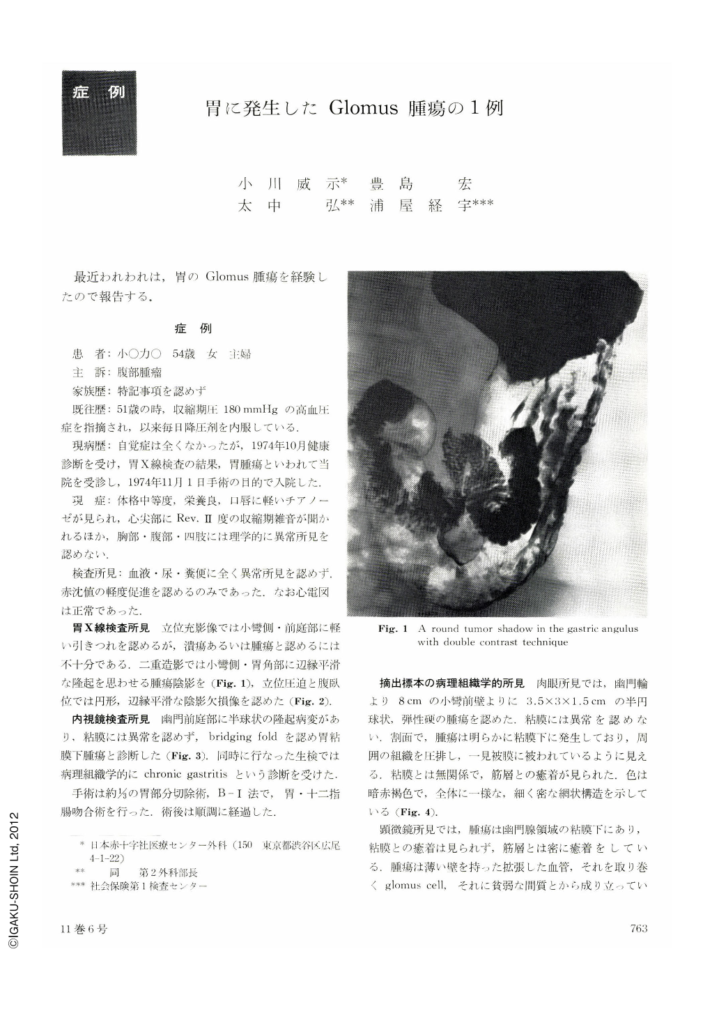
胃X線検査所見 立位充影像では小彎側・前庭部に軽い引きつれを認めるが,潰瘍あるいは腫瘍と認めるには不十分である.二重造影では小彎側・胃角部に辺縁平滑な隆起を思わせる腫瘍陰影を(Fig. 1),立位圧迫と腹臥位では円形,辺縁平滑な陰影欠損像を認めた(Fig. 2).
The case is a 54-year-old woman with glomus tumor of the stomach. She had a history of hypertension, but there were no abdominal symptoms. She visited our center because of a gastric tumor found by a medical routine check-up. After gastrofluoroscopy, endoscopy and histopathological examinations, she was diagnosed as harboring submucosal tumor of the stomach and underwent the operation. In the pyloric submucosal region, there was a walnut-sized tumor, which was demonstrated to be glomus tumor by the histopathological study. The tumor consisted of swollen blood vessel lumen and many glomus cells. According to the classification by Masson, this case is considered to belong to the complex type of the angiomatous type and epithelioid type.
From the viewpoint that the glomus tumor is benign, local gastric resection rather than partial resection should be performed. However, since glomus tumor shows pseudoinfiltration and recurrence is possible by the remaining cells, wide resection is nesessary.
Since this case was preoperatively diagnosed simply as submucosal tumor of the stomach and it was not clear whether it was benign or malignant, partial gastric resection with anastomosis by B-I method was performed.

Copyright © 1976, Igaku-Shoin Ltd. All rights reserved.


