Japanese
English
- 有料閲覧
- Abstract 文献概要
- 1ページ目 Look Inside
- サイト内被引用 Cited by
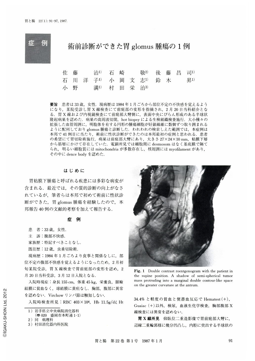
要旨 患者は33歳,女性.現病歴は1984年1月ごろから部位不定の不快感を覚えるようになり,某院受診し胃X線検査にて前庭部の変形を指摘され,2月20日当科紹介となる.胃X線および内視鏡検査にて前庭部大彎側に,表面中央にびらん形成のある半球状隆起病巣を認めた.病巣の高周波切開,hot biopsyによる生検組織検査施行.大小種々の拡張した血管周囲に,明胞体を有する円形の腫瘍細胞が好銀線維に数個ずつ取り囲まれるように配列しておりglomus腫瘍と診断した.われわれの検索しえた範囲では,本症例は本邦で41例目に当たり,術前に性状診断ができたのは本邦最初の症例と思われる.患者の希望にて胃切除術施行,病巣は前庭部大彎にあり,大きさ27×24×10mm,粘膜下層から筋層にかけて存在していた.電顕所見では細胞間にdesmosomはなく基底膜で隔てられ,明るい細胞質にはmitochondriaが多数存在し,核周囲にはmyofilamentがあり,その中にdence bodyを認めた.
The case is of a 33 year-old woman. From around January, 1984, she began to feel discomfort with no definite site indicated. Because of deformation of the antrum discovered in stomach x-ray examinations at a certain clinic, she was referred to this hospital. Stomach x-ray and endoscopic examination revealed a semispherical elevated lesion with erosion on its surface on the side of greater curvature at the antrum. Histologic examination of the lesion by high frequency incision and hot biopsy revealed arrangement of round tumor cells with clear cytoplasm, several of which were enclosed by reticular fiber around dilated blood vessels, small and large. The case was diagnosed as glomus tumor.
As far as we can ascertain, this is the 41st case in Japan and apparently it is the first case in Japan, of which normal diagnosis could be made preoperatively. At the request of the patient, gastrectomy was performed. The lesion was located at the antrum on the greater curvature; it measured 27×24×10 mm, extending from the lower layer of the mucosa to tunica muscularis. Electron microscopic examinations revealed no desmosomes between cells, but many mitochondria in the clear cytoplasm separated by the basement membrane and myofilament containing dense bodies.

Copyright © 1987, Igaku-Shoin Ltd. All rights reserved.


