Japanese
English
- 有料閲覧
- Abstract 文献概要
- 1ページ目 Look Inside
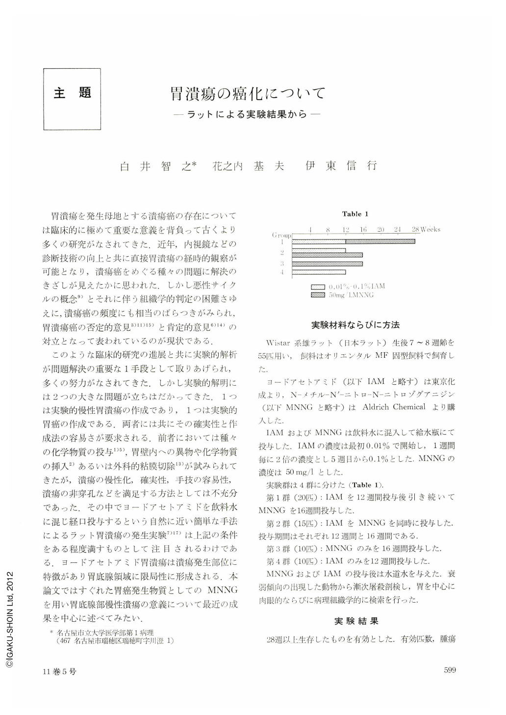
胃潰瘍を発生母地とする潰瘍癌の存在については臨床的に極めて重要な意義を背負って古くより多くの研究がなされてきた.近年,内視鏡などの診断技術の向上と共に直接胃潰瘍の経時的観察が可能となり,潰瘍癌をめぐる種々の問題に解決のきざしが見えたかに思われた.しかし悪性サイクルの概念9)とそれに伴う組織学的判定の困難さゆえに,潰瘍癌の頻度にも相当のばらつきがみられ,胃潰瘍癌の否定的意見3)11)15)と肯定的意見6)14)の対立となって表われているのが現状である.
このような臨床的研究の進展と共に実験的解析が問題解決の重要な1手段として取りあげられ,多くの努力がなされてきた.しかし実験的解明には2つの大きな問題が立ちはだかってきた.1つは実験的慢性胃潰瘍の作成であり,1つは実験的胃癌の作成である.両者には共にその確実性と作成法の容易さが要求される.前者においては種々の化学物質の投与1)5),胃壁内への異物や化学物質の挿入2)あるいは外科的粘膜切除13)が試みられてきたが,潰瘍の慢性化,確実性,手技の容易性,潰瘍の非穿孔などを満足する方法としては不充分であった.その中でヨードアセトアミドを飲料水に混じ経口投与するという自然に近い簡単な手法によるラット胃潰瘍の発生実験7)17)は上記の条件をある程度満すものとして注目されるわけである.ヨードアセトアミド胃潰瘍は潰瘍発生部位に特徴があり胃底腺領域に限局性に形成される.本論文ではすぐれた胃癌発生物質としてのMNNGを用い胃底腺部慢性潰瘍の意義について最近の成果を中心に述べてみたい.
The effect of ulcers induced by iodoacetamide on the development of gastric tumors by N-methyl-N'-nitro-N-nitrosoguanidine (MNNG) was studied in male Wistar strain rats. Animals were divided into 4 experimental groups and treated as follows: Group 1 was given 0.1% iodoacetamide (IAM) in the drinking water for 12 weeks and then 50 mg MNNG/ liter solution to drink for 16 weeks. Group 2 was given MNNG solution for 16 weeks with IAM for the first 12 weeks. Group 3 was treated with MNNG alone for 16 weeks. Group 4 was treated with IAM alone for 12 weeks. Animals in the groups 1 and 2 treated with IAM and MNNG developed a high incidence of tumors including adenocarcinoma in the fundic region, and no fundic tumors were seen in group 3. There was a significant difference (p<0.05) in the incidence of the fundic adenocarcinomas between groups 1 and 2 and group 3, but not in that incidence between groups 1 and 2. Animals in the groups 1 and 2 developed adenocarcinomas in the pyloric region as well as in the group 3. One of the fundic adenocarcinomas was poorly-differentiated type with loss of glandular arrangement, but others were well differentiated type. The tumors in the fundic region were located in the region where ulcers were induced by IAM, and especially, fundic adenocarcinomas were located on and/or in the scar tissue of the ulcer and were invasive. IAM induced the ulcers in the fundic region along the limiting ridge of the rat stomach and did not give any histopathological changes in both of the area of the fundic region and the pyloric region. Regenerating epithelium on areas of ulcer differentiated progressively, and the renewed mucosa showed pyloric metaplasia. It was concluded from these findings that the regenerated mucosa in an ulcer was capable of responding to carcinogenic stimulation and carcinomas arose from it finally.

Copyright © 1976, Igaku-Shoin Ltd. All rights reserved.


