Japanese
English
- 有料閲覧
- Abstract 文献概要
- 1ページ目 Look Inside
- サイト内被引用 Cited by
胃の類腺癌は極めて稀とされ,福田ら36)によれ
ば内外文献を通じて72例,うち本邦例は22例にすぎなかったという.また福田36),中村31),緒方33),佐野34),鮫島35),桑原37),松永38)らの例を加えると本邦例は40例となり,早期胃癌だけに限ってみると,鮫島らの報告した1例だけである.われわれも最近,類腺癌の早期胃癌例を経験した.本例は早期胃癌例として第2例目であり,また術前の内視鏡検査によって類腺癌と診断された最初の例となる.しかも本例は病理組織学的に多彩な所見を示し,発生論的にも興味ある症例と思われるので報告する.
症例
患 者:72歳女子
主 訴:全身倦怠感,食後の胃部不快感
家族歴:両親ともすでに失っているが,生前は比較的健康であつた.兄弟および子供は健在で,胃疾患,胃症状をもつものはいない.
既往歴:50年来,ときどき食後に胃部不快感と軽い心窩部痛をおぼえることがあったが,とくに治癒をうけるほどではなかった.
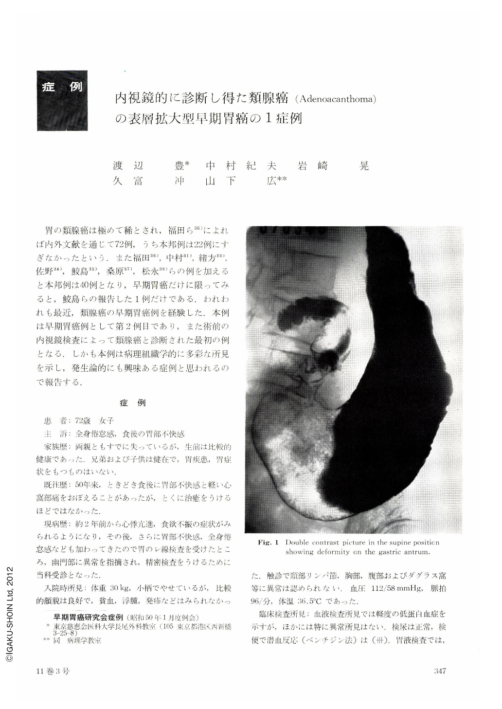
現病歴:約2年前から心悸亢進,食欲不振の症状がみられるようになり,その後,さらに胃部不快感,全身倦怠感なども加わってきたので胃のレ線検査を受けたところ,幽門部に異常を指摘され,精密検査をうけるために当科受診となった.
A report is made of early gastric acanthoma that showed interesting histopathologic changes.
Adenoacanthoma of the stomach is deemed a neoplasm of rare occurrence. Review of the literature shows that in Japan there have been only 36 reported cases of this growth including a single instance in early cancer stage. The present case is the second report of it in Japan and the first case preoperatively diagnosed as such by endoscopy.
The patient, a 72-year-old woman had chief complaints of malaise and postprandial unpleasant sensation of the stomach. Chief changes shown by x-ray were a shadow defect in the anterior part of the pyloric region and stiffening of the pyloric lesser curvature. Subseqnent endoscopy revealed Ⅱc of superficial spreading type in the entire pyloric region with partial Ⅱa-like changes, so that a diagnosis of early gastric cancer was first made. At the same time a submucosal tumor was seen on the anterior wall of the upper part of the body. As biopsy of the pyloric region revealed squamous cells and signet ring cells, the diagnosis was then adenoacanthoma.
Special features of the resected stomach were (1) superficial spreading type of sm early cancer stretching from the whole pyloric region up to the lower part of the body; (2) the majority of the lesion was poorly differentiated adenocarcinoma studded with differentiated squamous cell carcinoma, which was at places hard to distinguish from non-cancerous squamous epithelium and showed at other places sweat-gland-like structure; and (3) a leiomyoma in the submucosa of non-cancerous segments of the mucosa along with aberrant fundic glands in the duodenal mucosa. Further, cancerous tubules with partial squamous metaplasia were recognized within the chief lesion. In view of this, we have assumed that the chief lesion be adenoacanthoma caused by squamous metaplasia of poorly differentiated adenocarcinoma.
Copyright © 1976, Igaku-Shoin Ltd. All rights reserved.