Japanese
English
- 有料閲覧
- Abstract 文献概要
- 1ページ目 Look Inside
膵臓癌の予後は近年における著しい診断技術面での進歩,外科治療面での進歩にもかかわらず,きわめて悲観的であり,いまだこの暗黒の大陸を早期癌として診断治療することは困難である.筆者らは分化の高い膵腺房細胞癌の1例に膵頭十二指腸切除を施行し,5年生存を得たのでここに報告する.著者らの渉猟しえた範囲では,本邦においての膵腺房細胞癌の5年生存例の報告は無く,欧米文献にもその長期生存例を見ない貴重なる症例と考えている.
症例
症 例:K. M(♀)36歳
家族歴:特記すべきものはない.
既往歴:18歳 急性虫垂炎虫垂電切除.酒少量,煙草20本/日.
現病歴:1969年1月初,右季肋部に鳩卵大の腫瘤に気づく.同年9月初,腫瘤全体の硬度は増し,同部位にとう痛が発現,近医を受診,胃X線検査にて異常を指摘された.同年10月4日当院外科受診.全経過を通じて,貧血,黄疸,体重減少は認められない.病悩期間10カ月.
現 症:体格中等度.黄疽,貧血を認めず.右季肋部に11.0×9.0cmの境界鮮明なる硬い表面平滑の腫瘤を触知,可動性はない.肝腫脹,脾腫脹,腹水,脾動静脈血管雑音,Courvoisier's signはいずれも認められない.
一般検査:ー般検査所見をTable 1に示す.血清アミラーゼ値の軽度上昇を認めたほか,特に異常を認めず,P. Sテストなど膵外分泌試験は施行していない.
Acinar cell carcinoma of the pancreas has been reported to be 13% of entire carcinoma of the pancreas by Miller (1951), although long-termed survival over 5 years is not yet reported, as far as authors reviewed. This is a case report on curative resection which was performed on one of 2 acinar cell carcinomas diagnosed by histological examination of 66 cases of pancreatic carcinoma.
Case : Thirty three year-old female without anemia, jaundice or weight loss, was admitted with chief complaint of abdominal mass. It was the size of a goose egg, immobile and located in the right hypochondrium. Duration of illness was 10 months.
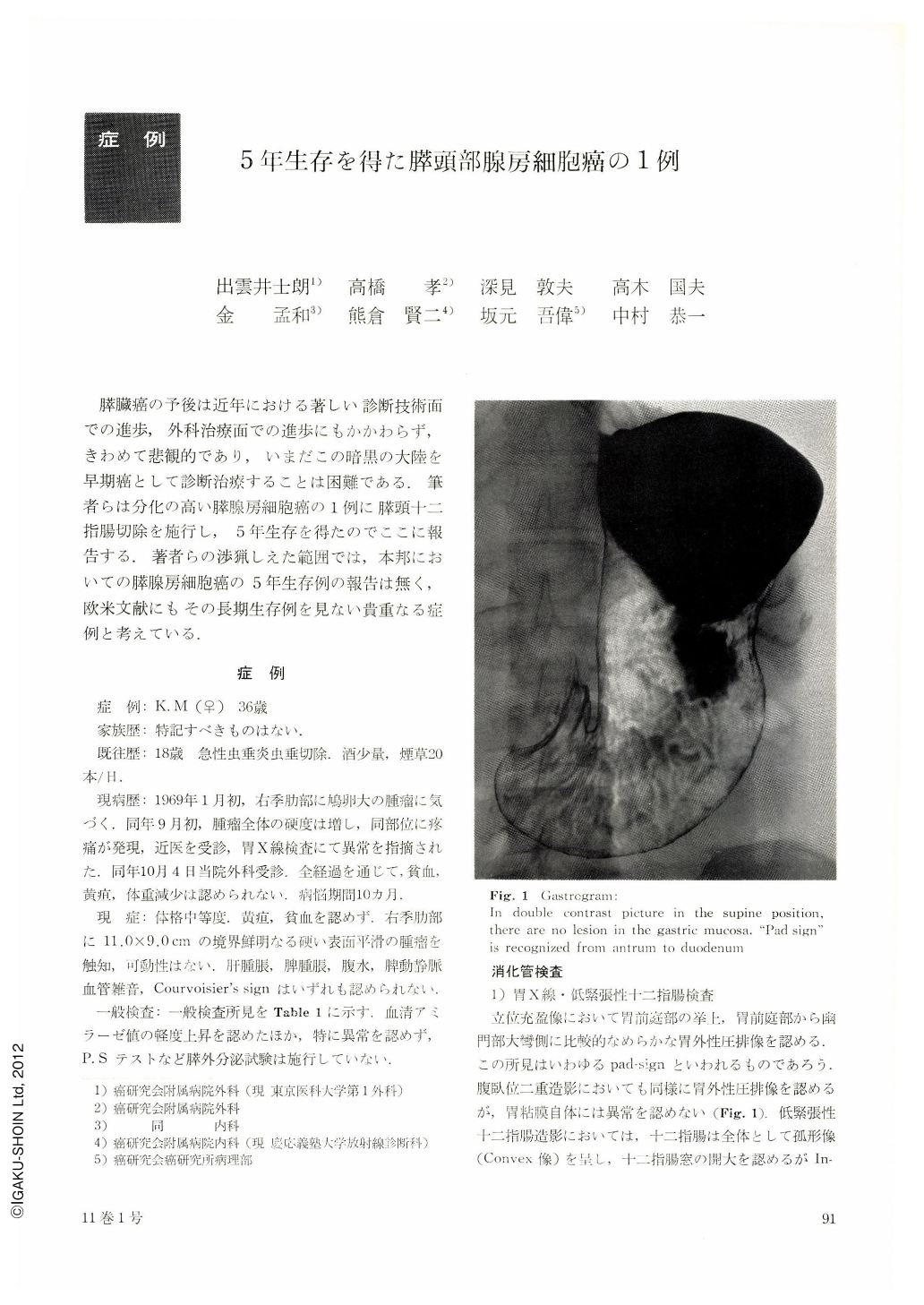
Gastrogram, hypotonic duodenography revealed convexity of the duodenum without Frostberg's phenomenon, irregularity of the duodenal mucosa relating to the extramural compression of tumor, without ulceration. Drop infusion cholecystocholangiogram demonstrated that the choledocus was not dilated but deviated laterally by tumor. Selective angiography revealed a space-occupying lesion showing cystic formationand highly developed vascularity. Coeliac angioscintinography (131 IMAA) suggested pancreas cyst.
Endoscopic examination of the duodenum documented that the mucosa of entire duodenum was free from lesion but endoscopic cannulation of papilla of Vater was impossible. Cytology of duodenal juice resulted in Class I. Pancreaticoduodenectomy was performed on 5 th Nov., 1969. Operative findings were that the large encapsulated tumor having abundant blood vessels 12×10×10 cm in size was located in the head of pancreas without any evidence of metastasis in the regional lymph nodes. Histological diagnosis was well differentiated acinar cell carcinoma of the pancreas. Atypism of the nuclei was not so prominent Tumor was encountered as a slow growing malignant one from the point of view of huge tumor and of partial invasion to capsule. Postoperative course was uneventful showing no evidence of metastasis or recurrence at the time of 5 years and 2 months after surgery.
Copyright © 1976, Igaku-Shoin Ltd. All rights reserved.