Japanese
English
- 有料閲覧
- Abstract 文献概要
- 1ページ目 Look Inside
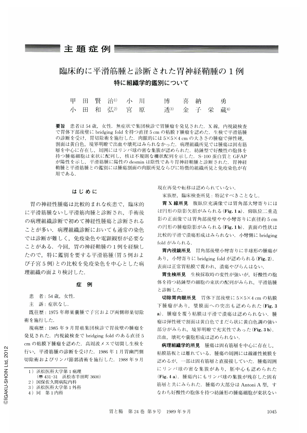
要旨 患者は54歳,女性.無症状で集団検診で胃腫瘤を発見された.X線,内視鏡検査で胃体下部後壁にbridging foldを持つ直径5cmの粘膜下腫瘤を認めた.生検で平滑筋腫の診断を受け,胃切除術を施行した.肉眼的には5×5×4cmの大きさの腫瘤で弾性硬,割面は黄白色,境界明瞭で出血や壊死はみられなかった.病理組織所見では腫瘍は固有筋層を中心に存在し,周囲にはリンパ球の密な集簇が認められた.紡錘型で好酸性の胞体を持つ腫瘍細胞は束状に配列し,核は不規則な柵状配列を示した.S-100蛋白質とGFAPが陽性を示し,平滑筋腫に陽性のdesminは陰性であり胃神経鞘腫と診断された.胃神経鞘腫と平滑筋腫との鑑別には腫瘍割面の肉眼所見ならびに特徴的組織所見と免疫染色が有用である.
A tumor of the stomach was detected in an asymptomatic 54-year-old woman during a mass survey. Endoscopic examination and radiography showed a submucosal tumor. Biopsy specimen was diagnosed as leiomyoma and gastrectomy was performed. The resected specimen showed a submucosal tumor, 5×5×4 cm in size, at the posterior wall of the lower corpus. The tumor was elastic firm and well-circumscribed and its cut surface was whitish and partly yellow. No ulcer, necrosis or hemorrhage was present.
Microscopically the tumor seemed to arise from the muscularis propria and was surrounded by lymphoid aggregates. The tumor was composed of bundles of spindle cells and their nuclei arranged in vague palisade. The nuclei revealed moderate variety in size and shape but mitoses were rare. Immunohistochemically S-100 protein and GFAP were positive but desmin was negative. Electron microscopy showed cytoplasmic extension with continuous basal lamina. There has been no recurrence for two years and eight months.
Neurilemoma of the stomach is rare and it is difficult to differentiate from leiomyoma not only clinically but pathologically in most cases. Characteristic patterns of nuclear and cytoplasmic arrangements and peritumoral lymphoid aggregates suggest that “neurilemoma” would be a correct diagnosis. Immunostaining for S-100 protein, GFAP and desmin is useful for this differentiation.

Copyright © 1989, Igaku-Shoin Ltd. All rights reserved.


