Japanese
English
- 有料閲覧
- Abstract 文献概要
- 1ページ目 Look Inside
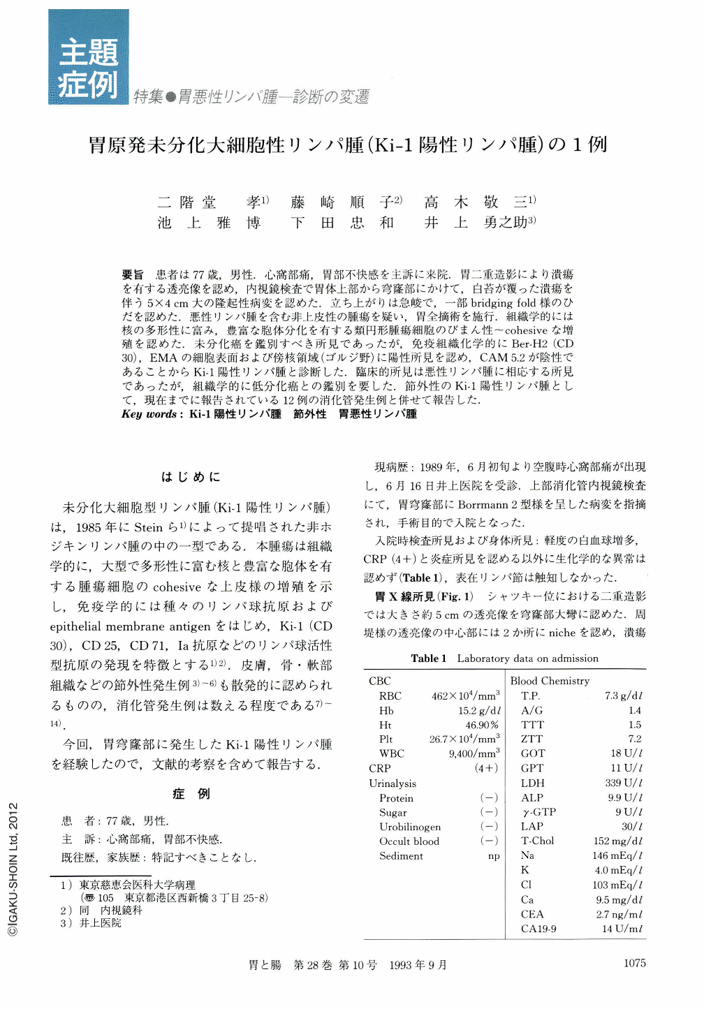
要旨 患者は77歳,男性.心窩部痛,胃部不快感を主訴に来院.胃二重造影により潰瘍を有する透亮像を認め,内視鏡検査で胃体上部から穹窿部にかけて,白苔が覆った潰瘍を伴う5×4cm大の隆起性病変を認めた.立ち上がりは急峻で,一部bridging fold様のひだを認めた.悪性リンパ腫を含む非上皮性の腫瘍を疑い,胃全摘術を施行.組織学的には核の多形性に富み,豊富な胞体分化を有する類円形腫瘍細胞のびまん性~cohesiveな増殖を認めた.未分化癌を鑑別すべき所見であったが,免疫組織化学的にBer-H2(CD30),EMAの細胞表面および傍核領域(ゴルジ野)に陽性所見を認め,CAM5.2が陰性であることからKi-1陽性リンパ腫と診断した.臨床的所見は悪性リンパ腫に相応する所見であったが,組織学的に低分化癌との鑑別を要した.節外性のKi-1陽性リンパ腫として,現在までに報告されている12例の消化管発生例と併せて報告した.
A case of a 77-year-old man with anaplastic large cell lymphoma of the stomach is reported. He was admitted complaining of epigastralgia. No superficial lymph nodes were palpable. Results of laboratory data on admission were unremarkable. Double contrast radiograph of the stomach demonstrated irregular and nodular elevation and irregular niche. Endoscopi cexamination demonstrated multiple white coating which seems to be adhesion, and surrounding elevation reveals a large nodule and has bridging fold. Clinical, radiological, and endoscopic findings were suggestive of non epithelial neoplasms including malignant lymphoma. Total gastrectomy was performed. Histological findings revealed diffuse and/or cohesive growth of the tumor cells with abundant cytoplasm and nuclear pleomorphism. Reed-Sternberg-like multi-nucleated giant cells were occasionally seen. It was difficult to distinguish between anaplastic carcinoma and non-Hodgkin's lymphoma in the histological findings. The tumor cells were immunoreactive with Ber-H2 (Ki-1, CD30) and EMA on cell surface and para-nuclear area. Cytokeratins, LCA, L26 (pan B), and Leu 22 (pan T) showed negative reaction.
This is an interesting case of extranodal anaplastic large-cell lymphoma. Ki-1 positive lymphomas of the gastro-intestinal tract are reviewed.

Copyright © 1993, Igaku-Shoin Ltd. All rights reserved.


