Japanese
English
- 有料閲覧
- Abstract 文献概要
- 1ページ目 Look Inside
- サイト内被引用 Cited by
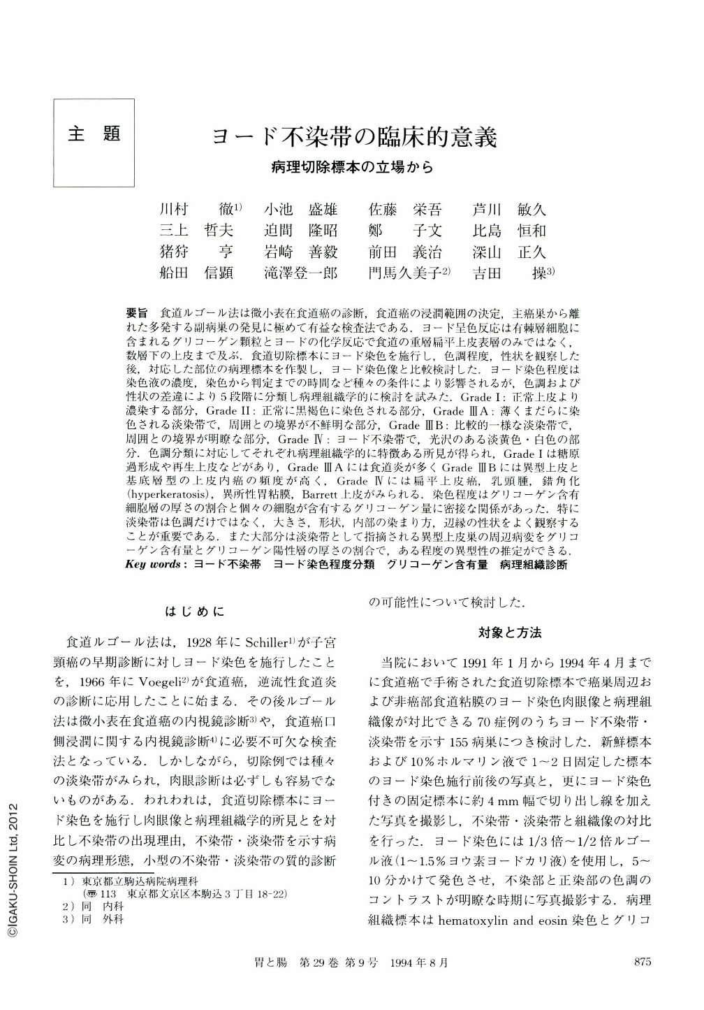
要旨 食道ルゴール法は微小表在食道癌の診断,食道癌の浸潤範囲の決定,主癌巣から離れた多発する副病巣の発見に極めて有益な検査法である.ヨード呈色反応は有棘層細胞に含まれるグリコーゲン顆粒とヨードの化学反応で食道の重層扁平上皮表層のみではなく,数層下の上皮まで及ぶ.食道切除標本にヨード染色を施行し,色調程度,性状を観察した後,対応した部位の病理標本を作製し,ヨード染色像と比較検討した.ヨード染色程度は染色液の濃度,染色から判定までの時間など種々の条件により影響されるが,色調および性状の差違により5段階に分類し病理組織学的に検討を試みた.Grade Ⅰ:正常上皮より濃染する部分,Grade Ⅱ:正常に黒褐色に染色される部分,Grade ⅢA:薄くまだらに染色される淡染帯で,周囲との境界が不鮮明な部分,Grade ⅢB:比較的一様な淡染帯で,周囲との境界が明瞭な部分,Grade Ⅳ:ヨード不染帯で,光沢のある淡黄色・白色の部分.色調分類に対応してそれぞれ病理組織学的に特徴ある所見が得られ,Grade Ⅰは糖原過形成や再生上皮などがあり,Grade ⅢAには食道炎が多ⅢBには異型上皮と基底層型の上皮内癌の頻度が高く,Grade Ⅳには扁平上皮癌,乳頭腫,錯角化(hyperkeratosis),異所性胃粘膜,Barrett上皮がみられる.染色程度はグリコーゲン含有細胞層の厚さの割合と個々の細胞が含有するグリコーゲン量に密接な関係があった.特に淡染帯は色調だけではなく,大きさ,形状,内部の染まり方,辺縁の性状をよく観察することが重要である.また大部分は淡染帯として指摘される異型上皮巣の周辺病変をグリコーゲン含有量とグリコーゲン陽性層の厚さの割合で,ある程度の異型性の推定ができる.
Lugol staining is a useful method to detect the small superficial carcinoma and to decide the proximal resection line of esophageal carcinoma with unexpectedly wide spread. To know the histopathological meaning of the Lugol unstained area of the esophagus, we examined 155 unstained lesions in resected esophageal specimens. Although the Lugol staining pattern is delicately different in each case due to the concentration of the iodine solution and the time needed to dye with good contrast, we classified the Lugol staining pattern into five grades by the staining grade and the marginal shape. Grade Ⅰ; hyperstained area. Grade Ⅱ; normal stained area. Grade ⅢA; less stained area with illdemarcated margin. Grade ⅢB; lesss tained area with well-demarcated margin. Grade Ⅳ; no stained area. We analyzed the relationship between grades and the histopathlogical findings by measuring the thickness of the glycogen-containing cell layers. We obtained the following conclusions.
1) Lugol test, which is a chemical reaction between glycogen and iodine, involves the prickle cell layer of the squamous epithelium.
2) The Lugol unstained areas correspond to such lesions as esophagitis, atypical epithelium, squamous cell carcinoma, so-called carcinosarcoma, Barrett esophagus, ectopic gastric mucosa, papilloma, and hyperkeratosis.
3) Staining grades correlate with the glycoge ncon tent in the epithelium and the ratio of the glycogen con-taining cell layers to the whole epithelium layers.
4) This classification of the Lugol staining pattern corresponds well to the histopathological findings.
5) Analysis of the ratio of glycogen containing cells might be helpful in differentiating esophagitis, atypical epithelium and carcinoma in situ.

Copyright © 1994, Igaku-Shoin Ltd. All rights reserved.


