Japanese
English
- 有料閲覧
- Abstract 文献概要
- 1ページ目 Look Inside
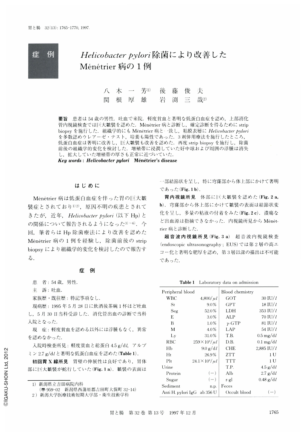
要旨 患者は54歳の男性.吐血で来院.軽度貧血と著明な低蛋白血症を認め,上部消化管内視鏡検査では巨大皺襞を認めた.Ménétrier病と診断し,確定診断を得るためにstrip biopsyを施行した.組織学的にもMénétrier病と一致し,粘膜表層にHelicobacter pyloriを多数認めウレアーゼ・テスト,培養も陽性であった.3剤併用療法を施行したところ,低蛋白血症は著明に改善し,巨大皺襞も改善を認めた.再度strip biopsyを施行し,除菌前後の組織学的変化を検討した.増殖帯に浸潤していた好中球および周囲の浮腫は消失し,拡大していた増殖帯の厚さも正常に近づいていた.
A 54-year-old man visited our hospital because he had hematomesis. Laboratory tests revealed anemia and low proteinemia. Initial endoscopy showed no bleeding focus, but enlarged folds and nodular surface of mucosa with rich mucus on the greater curvature of the corpus were noticed. A barium enema study of the upper gastrointestinal tract showed enlarged gastric folds of the body. He was diagnosed as having Ménétrier's disease. To make an accurate diagnosis histologically, strip biopsy of an enlarged gastric fold was carried out and this histological finding coincided with the features of Ménétrier's disease. There were also abundant H. pylori-like-organisms on the mucus and the surface epithelia. The patient was treated with lansoprazole (30 mg), amoxicillin (1,000 mg) and clarithromycin (600 mg) for 14 days. The levels of total protein and serum albumin returned to normal 9 weeks after eradication. Strip biopsy was again carried out on the same part of the stomach 16 weeks after eradication. At this time, endoscopic finding showed improvement of enlarged giant folds. Histological study after eradication revealed the following results: Hypertrophy and corkscrew appearance of surface epithelia had disappeared. Disappearance of infiltration of inflammatory cells and edema made arrangement of glands closen. The cystic dilatation of glands decreased. The patient has been clinically well during the 22 months since eradication therapy was terminated.

Copyright © 1997, Igaku-Shoin Ltd. All rights reserved.


